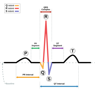
Electrocardiography is the process of recording the electrical activity of the heart over a period of time using electrodes placed over the skin. These electrodes detect the tiny electrical changes on the skin that arise from the heart muscle's electrophysiologic pattern of depolarizing and repolarizing during each heartbeat. It is very commonly performed to detect any cardiac problems.

Ventricular fibrillation is when the heart quivers instead of pumping due to disorganized electrical activity in the ventricles. It is a type of cardiac arrhythmia. Ventricular fibrillation results in cardiac arrest with loss of consciousness and no pulse. This is followed by death in the absence of treatment. Ventricular fibrillation is found initially in about 10% of people in cardiac arrest.

The systole is the part of the cardiac cycle during which some chambers of the heart muscle contract after refilling with blood. The term "systole" originates from New Latin via Ancient Greek συστολή (sustolē): from συστέλλειν via [σύν + στέλλειν. The use of systole, "to contract", is very similar to the use of the English term "to squeeze".

A ventricle is one of two large chambers toward the bottom of the heart that collect and expel blood received from an atrium towards the peripheral beds within the body and lungs. The atrium primes the pump. Interventricular means between the ventricles, while intraventricular means within one ventricle.
An ejection fraction (EF) is the volumetric fraction of fluid ejected from a chamber with each contraction. It can refer to the cardiac atrium, ventricle, gall bladder, or leg veins, although if unspecified it usually refers to the left ventricle of the heart. EF is widely used as a measure of the pumping efficiency of the heart and is used to classify heart failure types. It is also used as an indicator of the severity of heart failure, although it has recognized limitations.

Arrhythmogenic right ventricular dysplasia (ARVD), or arrhythmogenic right ventricular cardiomyopathy (ARVC), is an inherited heart disease.

Ventricular tachycardia is a type of regular, fast heart rate that arises from improper electrical activity in the ventricles of the heart. Although a few seconds may not result in problems, longer periods are dangerous. Short periods may occur without symptoms, or present with lightheadedness, palpitations, or chest pain. Ventricular tachycardia may result in cardiac arrest and turn into ventricular fibrillation. It is found initially in about 7% of people in cardiac arrest.

A ventricular septal defect (VSD) is a defect in the ventricular septum, the wall dividing the left and right ventricles of the heart. The extent of the opening may vary from pin size to complete absence of the ventricular septum, creating one common ventricle. The ventricular septum consists of an inferior muscular and superior membranous portion and is extensively innervated with conducting cardiomyocytes.

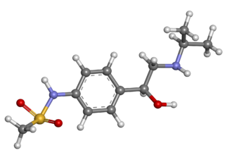
Sotalol is a medication used to treat abnormal heart rhythms. The U.S. Food and Drug Administration (FDA) advises that sotalol only be used for serious abnormal heart rhythms, because its prolongation of the QT interval carries a small risk of life-threatening polymorphic ventricular tachycardia known as torsade de pointes.

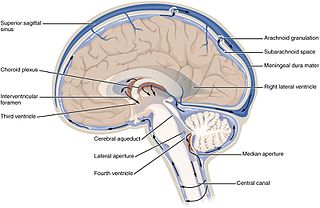
The lateral ventricles are the two largest cavities of the ventricular system of the human brain and contain cerebrospinal fluid (CSF). Each cerebral hemisphere contains a lateral ventricle, known as the left or right ventricle, respectively.

In the brain, the interventricular foramina are channels that connect the paired lateral ventricles with the third ventricle at the midline of the brain. As channels, they allow cerebrospinal fluid (CSF) produced in the lateral ventricles to reach the third ventricle and then the rest of the brain's ventricular system. The walls of the interventricular foramina also contain choroid plexus, a specialized CSF-producing structure, that is continuous with that of the lateral and third ventricles above and below it.
A transthoracic echocardiogram (TTE) is the most common type of echocardiogram, which is a still or moving image of the internal parts of the heart using ultrasound. In this case, the probe is placed on the chest or abdomen of the subject to get various views of the heart. It is used as a non-invasive assessment of the overall health of the heart, including a patient's heart valves and degree of heart muscle contraction. The images are displayed on a monitor for real-time viewing and then recorded.

The cardiac cycle is the performance of the human heart from the beginning of one heartbeat to the beginning of the next. It consists of two periods: one during which the heart muscle relaxes and refills with blood, called diastole, followed by a period of robust contraction and pumping of blood, dubbed systole. After emptying, the heart immediately relaxes and expands to receive another influx of blood returning from the lungs and other systems of the body, before again contracting to pump blood to the lungs and those systems. A normally performing heart must be fully expanded before it can efficiently pump again. Assuming a healthy heart and a typical rate of 70 to 75 beats per minute, each cardiac cycle, or heartbeat, takes about 0.8 seconds to complete the cycle.
A ventricular outflow tract is a portion of either the left ventricle or right ventricle of the heart through which blood passes in order to enter the great arteries.

Desmoplakin is a protein in humans that is encoded by the DSP gene. Desmoplakin is a critical component of desmosome structures in cardiac muscle and epidermal cells, which function to maintain the structural integrity at adjacent cell contacts. In cardiac muscle, desmoplakin is localized to intercalated discs which mechanically couple cardiac cells to function in a coordinated syncytial structure. Mutations in desmoplakin have been shown to play a role in dilated cardiomyopathy, arrhythmogenic right ventricular cardiomyopathy, striate palmoplantar keratoderma, Carvajal syndrome and paraneoplastic pemphigus.

Myosin essential light chain (ELC), ventricular/cardiac isoform is a protein that in humans is encoded by the MYL3 gene. This cardiac ventricular/slow skeletal ELC isoform is distinct from that expressed in fast skeletal muscle (MYL1) and cardiac atrial muscle (MYL4). Ventricular ELC is part of the myosin molecule and is important in modulating cardiac muscle contraction.

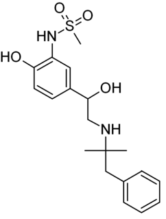
Zinterol is a beta-adrenergic agonist.
In electrocardiography, a strain pattern is a well-recognized marker for the presence of anatomic left ventricular hypertrophy (LVH) in the form of ST depression and T wave inversion on a resting ECG. It is an abnormality of repolarization and it has been associated with an adverse prognosis in a variety heart disease patients. It has been important in refining the role of ECG LVH criteria in cardiac risk stratification. It is thought that a strain pattern could also reflect underlying coronary heart disease.
Crista supraventricularis is a muscular ridge within the right ventricle of the heart. It is located between the tricuspid and pulmonic valves, at the junction of the right ventricular anterior (free) wall and the interventricular septum. It has a "U-shaped" morphology, which serves as a "trough" for the proximal right coronary artery.


