
Cerebrospinal fluid (CSF) is a clear, colorless body fluid found within the tissue that surrounds the brain and spinal cord of all vertebrates.

The abducens nerve or abducent nerve, also known as the sixth cranial nerve, cranial nerve VI, or simply CN VI, is a cranial nerve in humans and various other animals that controls the movement of the lateral rectus muscle, one of the extraocular muscles responsible for outward gaze. It is a somatic efferent nerve.

A heart valve is a one-way valve that allows blood to flow in one direction through the chambers of the heart. Four valves are usually present in a mammalian heart and together they determine the pathway of blood flow through the heart. A heart valve opens or closes according to differential blood pressure on each side.
The ventricular system is a set of four interconnected cavities known as cerebral ventricles in the brain. Within each ventricle is a region of choroid plexus which produces the circulating cerebrospinal fluid (CSF). The ventricular system is continuous with the central canal of the spinal cord from the fourth ventricle, allowing for the flow of CSF to circulate.

Pia mater, often referred to as simply the pia, is the delicate innermost layer of the meninges, the membranes surrounding the brain and spinal cord. Pia mater is medieval Latin meaning "tender mother". The other two meningeal membranes are the dura mater and the arachnoid mater. Both the pia and arachnoid mater are derivatives of the neural crest while the dura is derived from embryonic mesoderm. The pia mater is a thin fibrous tissue that is permeable to water and small solutes. The pia mater allows blood vessels to pass through and nourish the brain. The perivascular space between blood vessels and pia mater is proposed to be part of a pseudolymphatic system for the brain. When the pia mater becomes irritated and inflamed the result is meningitis.

The midbrain or mesencephalon is the rostral-most portion of the brainstem connecting the diencephalon and cerebrum with the pons. It consists of the cerebral peduncles, tegmentum, and tectum.

The fourth ventricle is one of the four connected fluid-filled cavities within the human brain. These cavities, known collectively as the ventricular system, consist of the left and right lateral ventricles, the third ventricle, and the fourth ventricle. The fourth ventricle extends from the cerebral aqueduct to the obex, and is filled with cerebrospinal fluid (CSF).
Hubert von Luschka, born Hubert Luschka, was a German anatomist. He lent his name to several structures, including the foramina of Luschka, Luschka's crypts, Luschka's joints, and Ducts of Luschka. His name is also associated with Luschka's law, an anatomical rule concerning location of the ureters.
In anatomy, Luschka's joints are formed between uncinate process or "uncus" below and uncovertebral articulation above. They are located in the cervical region of the vertebral column from C3 to C7. Two lips project upward from the superior surface of the vertebral body below, and one projects downward from the inferior surface of vertebral body above. They allow for flexion and extension and limit lateral flexion in the cervical spine.
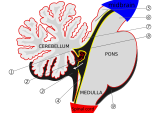
The median aperture drains cerebrospinal fluid (CSF) from the fourth ventricle into the cisterna magna. The two other openings of the fourth ventricle are the lateral apertures, one on the left and one on the right, which drain cerebrospinal fluid into the cerebellopontine angle cistern. The median foramen on axial images is posterior to the pons and anterior to the caudal cerebellum. It is surrounded by the obex and gracile tubercles of the medulla, tela choroidea of the fourth ventricle and its choroid plexus, which is attached to the cerebellar vermis.
The aperture of an optical system is the opening that limits the amount of light that can pass through.

In the brain, the interventricular foramina are channels that connect the paired lateral ventricles with the third ventricle at the midline of the brain. As channels, they allow cerebrospinal fluid (CSF) produced in the lateral ventricles to reach the third ventricle and then the rest of the brain's ventricular system. The walls of the interventricular foramina also contain choroid plexus, a specialized CSF-producing structure, that is continuous with that of the lateral and third ventricles above and below it.

The subarachnoid cisterns are spaces formed by openings in the subarachnoid space, an anatomic space in the meninges of the brain. The space is situated between the two meninges, the arachnoid mater and the pia mater. These cisterns are filled with cerebrospinal fluid (CSF).
The cisterna magna is one of three principal openings in the subarachnoid space between the arachnoid and pia mater layers of the meninges surrounding the brain. The openings are collectively referred to as the subarachnoid cisterns. The cisterna magna is located between the cerebellum and the dorsal surface of the medulla oblongata. Cerebrospinal fluid produced in the fourth ventricle drains into the cisterna magna via the lateral apertures and median aperture.

Vincent Alexander Bochdalek was a Bohemian anatomist and pathologist. His first name has also been given as Vincenc and Vincenz. Bochdalek was elected as member of the German Academy of Sciences Leopoldina.

The trigeminal motor nucleus contains motor neurons that innervate muscles of the first branchial arch, namely the muscles of mastication, the tensor tympani, tensor veli palatini, mylohyoid, and anterior belly of the digastric. It is situated in the upper pons, inferior to the lateral part of the floor of the fourth ventricle.

The tela choroidea is a region of meningeal pia mater that adheres to the underlying ependyma, and gives rise to the choroid plexus in each of the brain’s four ventricles. Tela is Latin for woven and is used to describe a web-like membrane or layer. The tela choroidea is a very thin part of the loose connective tissue of pia mater overlying and closely adhering to the ependyma. It has a rich blood supply. The ependyma and vascular pia mater – the tela choroidea, form regions of minute projections known as a choroid plexus that projects into each ventricle. The choroid plexus produces most of the cerebrospinal fluid of the central nervous system that circulates through the ventricles of the brain, the central canal of the spinal cord, and the subarachnoid space. The tela choroidea in the ventricles forms from different parts of the roof plate in the development of the embryo.
The pontine cistern is a subarachnoid cistern in the interval between ventral aspect of the pons, and the clivus. It contains the basilar artery. The lateral aperture opens into the pontine cistern just posterior to the cranial nerve VIII.
The lateral recess is a narrow extension of the fourth ventricle on either side projecting laterally posterior and around the inferior cerebellar peduncle, opening into the subarachnoid space at its lateral extremity as the lateral aperture to allow for the passage of cerebrospinal fluid.
The superior cistern is a dilation as a subarachnoid cistern of the subarachnoid space around the brain. It lies between the splenium of the corpus callosum and the superior surface of the cerebellum. It extends between the layers of the tela choroidea of the third ventricle. It contains the great cerebral vein, posterior cerebral artery, quadrigeminal artery, glossopharyngeal nerve, and the pineal gland.

