Syndecan

Last updated
Syndecan
PDB 1ejq EBI.jpg
Solution structure of the Syndecan-4 whole cytoplasmic domain in the presence of phosphatidylinositol 4,5-bisphosphate.
Identifiers
SymbolSyndecan
Pfam PF01034
InterPro IPR001050
PROSITE PDOC00745
SCOP2 1ejp / SCOPe / SUPFAM
OPM superfamily 535
OPM protein 6ith
Membranome 18
Available protein structures:
Pfam   structures / ECOD  
PDB RCSB PDB; PDBe; PDBj
PDBsum structure summary

Syndecans are single transmembrane domain proteins that are thought to act as coreceptors, especially for G protein-coupled receptors. More specifically, these core proteins carry three to five heparan sulfate and chondroitin sulfate chains, i.e. they are proteoglycans, which allow for interaction with a large variety of ligands including fibroblast growth factors, vascular endothelial growth factor, transforming growth factor-beta, fibronectin and antithrombin-1. Interactions between fibronectin and some syndecans can be modulated by the extracellular matrix protein tenascin C.

Contents

Family members and Structure

The syndecan protein family has four members. Syndecans 1 and 3 and syndecans 2 and 4, making up separate subfamilies, arose by gene duplication and divergent evolution from a single ancestral gene. [1] The syndecan numbers reflect the order in which the cDNAs for each family member were cloned. All syndecans have an N-terminal signal peptide, an ectodomain, a single hydrophobic transmembrane domain, and a short C-terminal cytoplasmic domain. [2] All syndecans are anchored to plasma membrane via a 24-25 amino acid long hydrophobic transmembrane domain, in contrast to another type of cell surface proteoglycans that attaches to cell membrane using a glycosyl-phosphatidyl-inositol linkage. [3] The most obvious differences between syndecans include (together with differences in distribution) the subclassification of the family depending on the existence of GAG binding sites either at both ends of the ectodomain (syndecan-1 and - 3) or at the distal part only (syndecan-2 and -4) and a relatively long Thr-Ser-Pro-rich area in the middle of syndecan- 3's ectodomain. [3] The ectodomains show the least amount of amino acid sequence conservation, not more than 10–20%; in contrast, the transmembrane and cytoplasmic domains share approximately 60–70% amino acid sequence identity. [4] The transmembrane domains contain an unusual alanine/glycine sequence motif, while the cytoplasmic domain is essentially composed of two regions of conserved amino acid sequence (C1 and C2), separated by a central variable sequence of amino acids that is distinct for each family member (V).

In mammalian cells, syndecans are expressed by unique genes located on different chromosomes. This is general lack of evidence of alternate splicing in syndecan genes. All members of the syndecan family have 5 exons. The difference in size of the syndecans is credited to the variable length of exon 3, which encodes a spacer domain [1, 14]. In humans, the amino acid length of syndecan 1, 2, 3 and 4 is 310, 201, 346 and 198 respectively. Glycosaminoglycan chains, a member of the heparan sulfate group, are an important component of syndecan and are responsible for a diverse set of syndecan functions. The addition of glycosaminoglycans to syndecan is controlled by a series of post- translation events. The preferential site for the addition of glycosaminoglycans is on a serine residue followed by glycine residue, where the linker is attached for the elongation of the glycosaminoglycans by α-N-acetylglucosaminyltransferase I [1]. The linker is composed of four saccharides, first one being xylose, which is an unusual sugar in a unique place, attached to serine of the protein core and sequentially followed by two galactose and a β-D-glucuronic acid [1, 12].

Expression

Syndecans are expressed on the cell surface in a cell-specific manner. For example, in mouse cells and tissues, syndecan 1 is highly expressed in fibroblastic and epithelial cells. It is especially high in keratinocytes whereas low in endothelial and neural cells. These tissues include skin, liver, kidney and lungs. Syndecan 2 is highly expressed in endothelial, neural, and fibroblastic cells, whereas it has low expression levels in epithelial cells. It is specific to tissues such as the liver, endothelia and fibroblasts. Syndecan 3 is highly expressed in neural cells, but has low or undetectable amount in epithelial cells. In tissues, it is specific to the brain and expressed at low levels in liver, kidney, lung and small intestine. Syndecan 4 is highly expressed by epithelial and fibroblastic cells, but has low expression levels in neural and endothelial cells. In tissues, it is preferentially expressed in the liver and lungs [11].

Functions

Functionality of syndecan is contributed by glycosaminoglycans which help in the interaction with different extracellular ligands. Depending upon the cellular localization of syndecan, glycosaminoglycans have different structures to accommodate the functional needs of the region. The syndecans are known to form homologous oligomers that may be important for their functions. [5]

Functions of syndecan can be categorized in four ways. First is growth-factor-receptor activation. Glycosaminoglycans attached to the syndecan help binding of the various growth factors for activation of important cellular signaling mechanisms. Growth factors such as FGF2, HGF, EGF, VEGF, neuregulins and others interact with syndecans [1, 2, 8]. For example, at the site of tissue injury, the soluble syndecan-1 ectodomains are cleaved by heparanases, producing heparin-like fragments that activate bFGF [13]. Whereas most growth factors interact with syndecans via heparan sulfate chains, the prosecretory mitogen lacritin requires heparanase to both expose and create a binding site in the N-terminus of syndecan 1. [6] [7]

Second is matrix adhesion. Syndecans bind to structural extracellular matrix molecules such as collagens I, III, V, fibronectin, thrombospondin, and tenascin to provide structural support for the adhesion [1, 2].

A third function is cell–cell adhesion. Evidence for syndecan's role in cell–cell adhesion comes from the human myeloma cell line. These myeloma cells had a deficiency in the ability to adhere to one another in a rotation-mediated aggregation matrix. This deficiency is attributed to the lack of syndecan 1 expression. Syndecan 4 also interacts with integrin proteins for cell–cell adhesion [1, 2, 12].

A final role is in tumor suppression and progression. Syndecans act as tumor inhibitors by preventing cellular proliferation of tumor cell lines. For example, in the epithelial-derived tumor cell line, S115, the syndecan 1 ectodomain suppresses the growth of S115 cells without affecting the growth of normal epithelial cells [7]. However, syndecan 1 expression also has a role in tumor progression in myeloma and other cancers [5, 6, 9, 15]. It associates with intracellular actin cytoskeleton and helps maintain normal epithelium sheet morphology

Protein Domains

The syndecan proteins can contain the following protein domains,

Clinical significance

Endometriosis

Syndecan-4 is upregulated in endometriosis and inhibition of syndecan-4 in human endometriotic cells results in a reduction of invasive growth in vitro and changes in matrix metalloproteinase expression. [10]

Osteoarthritis

Syndecan-4 is upregulated in osteoarthritis and inhibition of syndecan-4 reduces cartilage destruction in mouse models of OA. [11]

Metabolic regulation and body composition

The Drosophila homologue dSdc and human SDC4 have been implicated in energy homeostasis. [12]

Multiple Myeloma

Syndecan1 is upregulated in multiple myeloma. High levels of shed syndecan1 in a patient's serum typically is correlated with poor prognosis.

Syndecan 1 is the most studied of all the syndecans in cancer research. Many studies have shown that syndecan 1 plays an important role in cancer progression, and also can be used as cancer biomarker. For example, syndecan 1 expression is higher in the bone marrow of the patients suffering from the multiple myeloma [9]. In one published study, the cells expressing the soluble syndecan 1 ectodomain promoted the growth and metastasis of B-lymphoid tumors more extensively than cells bearing surface syndecan 1 or lacking syndecan 1 expression [16]. Similarly, syndecan 1 expression has been linked with low differentiation in squamous cell carcinoma of the head and neck [15].

Syndecan 1 also has been linked with cancer progression by mediating the effects of growth factors in the cells. For example, syndecan 1 expression is increased in ductal breast carcinomas and is associated with factors of angiogenesis and lymphangiogenesis [5]. Studies from patients suffering from endometrial cancer have shown that these patients have increased syndecan 1 expression, and also that expression of this protein positively regulates the endometrial hyperplasia that can progress to endometrial cancer [6].

Related Research Articles

<span class="mw-page-title-main">Glycosaminoglycan</span> Polysaccharides found in animal tissue

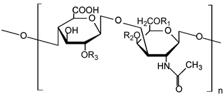
Glycosaminoglycans (GAGs) or mucopolysaccharides are long, linear polysaccharides consisting of repeating disaccharide units. The repeating two-sugar unit consists of a uronic sugar and an amino sugar, except in the case of the sulfated glycosaminoglycan keratan, where, in place of the uronic sugar there is a galactose unit. GAGs are found in vertebrates, invertebrates and bacteria. Because GAGs are highly polar molecules and attract water; the body uses them as lubricants or shock absorbers.

<span class="mw-page-title-main">P-selectin</span> Type-1 transmembrane protein

P-selectin is a type-1 transmembrane protein that in humans is encoded by the SELP gene.

<span class="mw-page-title-main">Versican</span> Protein-coding gene in the species Homo sapiens

Versican is a large extracellular matrix proteoglycan that is present in a variety of human tissues. It is encoded by the VCAN gene.

<span class="mw-page-title-main">Perlecan</span>

Perlecan (PLC) also known as basement membrane-specific heparan sulfate proteoglycan core protein (HSPG) or heparan sulfate proteoglycan 2 (HSPG2), is a protein that in humans is encoded by the HSPG2 gene. The HSPG2 gene codes for a 4,391 amino acid protein with a molecular weight of 468,829. It is one of the largest known proteins. The name perlecan comes from its appearance as a "string of pearls" in rotary shadowed images.

<span class="mw-page-title-main">Pleiotrophin</span> Protein in humans

Pleiotrophin (PTN) also known as heparin-binding brain mitogen (HBBM) or heparin-binding growth factor 8 (HBGF-8) or neurite growth-promoting factor 1 (NEGF1) or heparin affinity regulatory peptide (HARP) or heparin binding growth associated molecule (HB-GAM) is a protein that in humans is encoded by the PTN gene. Pleiotrophin is an 18-kDa growth factor that has a high affinity for heparin. It is structurally related to midkine and retinoic acid induced heparin-binding protein.

<span class="mw-page-title-main">Heparan sulfate</span> Macromolecule

Heparan sulfate (HS) is a linear polysaccharide found in all animal tissues. It occurs as a proteoglycan in which two or three HS chains are attached in close proximity to cell surface or extracellular matrix proteins. In this form, HS binds to a variety of protein ligands, including Wnt, and regulates a wide range of biological activities, including developmental processes, angiogenesis, blood coagulation, abolishing detachment activity by GrB, and tumour metastasis. HS has also been shown to serve as cellular receptor for a number of viruses, including the respiratory syncytial virus. One study suggests that cellular heparan sulfate has a role in SARS-CoV-2 Infection, particularly when the virus attaches with ACE2.

<span class="mw-page-title-main">Lacritin</span>

Lacritin is a 12.3 kDa glycoprotein encoded in humans by the LACRT gene. Lacritin's discovery emerged from a screen for factors that stimulate tear protein secretion. Lacritin is a secreted protein found in tears and saliva. Lacritin also promotes tear secretion, the proliferation and survival of epithelial cells, and corneal wound healing Lacritin is thus a multifunctional prosecretory mitogen with cell survival activity. Natural or bacterial cleavage of lacritin releases a C-terminal fragment that is bactericidal.

<span class="mw-page-title-main">TGFBR3</span> Protein-coding gene in the species Homo sapiens

Betaglycan also known as Transforming growth factor beta receptor III (TGFBR3), is a cell-surface chondroitin sulfate / heparan sulfate proteoglycan >300 kDa in molecular weight. Betaglycan binds to various members of the TGF-beta superfamily of ligands via its core protein, and bFGF via its heparan sulfate chains. TGFBR3 is the most widely expressed type of TGF-beta receptor. Its affinity towards all individual isoforms of TGF-beta is similarly high and therefore it plays an important role as a coreceptor mediating the binding of TGF-beta to its other receptors - specifically TGFBR2. The intrinsic kinase activity of this receptor has not yet been described. In regard of TGF-beta signalling it is generally considered a non-signaling receptor or a coreceptor. By binding to various member of the TGF-beta superfamily at the cell surface it acts as a reservoir of TGF-beta.

<span class="mw-page-title-main">Syndecan 1</span> Protein which in humans is encoded by the SDC1 gene

Syndecan 1 is a protein which in humans is encoded by the SDC1 gene. The protein is a transmembrane heparan sulfate proteoglycan and is a member of the syndecan proteoglycan family. The syndecan-1 protein functions as an integral membrane protein and participates in cell proliferation, cell migration and cell-matrix interactions via its receptor for extracellular matrix proteins. Syndecan-1 is a sponge for growth factors and chemokines, with binding largely via heparan sulfate chains. The syndecans mediate cell binding, cell signaling, and cytoskeletal organization and syndecan receptors are required for internalization of the HIV-1 tat protein.

<span class="mw-page-title-main">Glypican</span>

Glypicans constitute one of the two major families of heparan sulfate proteoglycans, with the other major family being syndecans. Six glypicans have been identified in mammals, and are referred to as GPC1 through GPC6. In Drosophila two glypicans have been identified, and these are referred to as dally and dally-like. One glypican has been identified in C. elegans. Glypicans seem to play a vital role in developmental morphogenesis, and have been suggested as regulators for the Wnt and Hedgehog cell signaling pathways. They have additionally been suggested as regulators for fibroblast growth factor and bone morphogenic protein signaling.

<span class="mw-page-title-main">Heparin-binding EGF-like growth factor</span> Protein-coding gene in the species Homo sapiens

Heparin-binding EGF-like growth factor (HB-EGF) is a member of the EGF family of proteins that in humans is encoded by the HBEGF gene.

<span class="mw-page-title-main">Syndecan-2</span> Protein-coding gene in the species Homo sapiens

Syndecan-2 is a protein that in humans is encoded by the SDC2 gene.

<span class="mw-page-title-main">Syndecan-4</span> Protein-coding gene in the species Homo sapiens

Syndecan-4 is a protein that in humans is encoded by the SDC4 gene. Syndecan-4 is one of the four vertebrate syndecans and has a molecular weight of ~20 kDa. Syndecans are the best-characterized plasma membrane proteoglycans. Their intracellular domain of membrane-spanning core protein interacts with actin cytoskeleton and signaling molecules in the cell cortex. Syndecans are normally found on the cell surface of fibroblasts and epithelial cells. Syndecans interact with fibronectin on the cell surface, cytoskeletal and signaling proteins inside the cell to modulate the function of integrin in cell-matrix adhesion. Also, syndecans bind to FGFs and bring them to the FGF receptor on the same cell. As a co-receptor or regulator, mutated certain proteoglycans could cause severe developmental defects, like disordered distribution or inactivation of signaling molecules.

<span class="mw-page-title-main">Heparanase</span> Mammalian protein found in Homo sapiens

Heparanase, also known as HPSE, is an enzyme that acts both at the cell-surface and within the extracellular matrix to degrade polymeric heparan sulfate molecules into shorter chain length oligosaccharides.

<span class="mw-page-title-main">Syndecan-3</span> Protein-coding gene in the species Homo sapiens

Syndecan-3 is a protein that in humans is encoded by the SDC3 gene.

<span class="mw-page-title-main">Glypican 1</span> Protein-coding gene in the species Homo sapiens

Glypican-1 (GPC1) is a protein that in humans is encoded by the GPC1 gene. GPC1 is encoded by human GPC1 gene located at 2q37.3. GPC1 contains 558 amino acids with three predicted heparan sulfate chains.

<span class="mw-page-title-main">NCR3</span>

Natural cytotoxicity triggering receptor 3 is a protein that in humans is encoded by the NCR3 gene. NCR3 has also been designated as CD337 and as NKp30. NCR3 belongs to the family of NCR membrane receptors together with NCR1 (NKp46) and NCR2 (NKp44).

<span class="mw-page-title-main">SULF1</span> Protein-coding gene in the species Homo sapiens

Sulfatase 1, also known as SULF1, is an enzyme which in humans is encoded by the SULF1 gene.

<span class="mw-page-title-main">TNFSF12-TNFSF13</span> Protein-coding gene in the species Homo sapiens

Tumor necrosis factor (ligand) superfamily, member 12-member 13, also known as TNFSF12-TNFSF13, is a human gene.

<span class="mw-page-title-main">Carbohydrate sulfotransferase</span>

Carbohydrate sulfotransferases are sulfotransferase enzymes that transfer sulfate to carbohydrate groups in glycoproteins and glycolipids. Carbohydrates are used by cells for a wide range of functions from structural purposes to extracellular communication. Carbohydrates are suitable for such a wide variety of functions due to the diversity in structure generated from monosaccharide composition, glycosidic linkage positions, chain branching, and covalent modification. Possible covalent modifications include acetylation, methylation, phosphorylation, and sulfation. Sulfation, performed by carbohydrate sulfotransferases, generates carbohydrate sulfate esters. These sulfate esters are only located extracellularly, whether through excretion into the extracellular matrix (ECM) or by presentation on the cell surface. As extracellular compounds, sulfated carbohydrates are mediators of intercellular communication, cellular adhesion, and ECM maintenance.

References

  1. Carey, D. J. (1997). "Syndecans: multifunctional cell-surface co-receptors". Biochem. J. 327 (Pt 1): 1–16. doi:10.1042/bj3270001. PMC   1218755 . PMID   9355727.
  2. Bernfield M, Kokenyesi R, et al. (1992). "Biology of syndecans: a family of transmembrane heparan sulfate proteoglycans". Annu. Rev. Cell Biol. 8: 365–393. doi:10.1146/annurev.cb.08.110192.002053. PMID   1335744.
  3. 1 2 Klaus Elenius & Markku Jalkanen (1994). "Function of the syndecans - a family of cell surface proteoglycans". Journal of Cell Science. 107 (11): 2975–2982. doi:10.1242/jcs.107.11.2975. PMID   7698997.
  4. David, G. (1 August 1993). "Integral membrane heparan sulfate proteoglycans". FASEB J. 7 (11): 1023–1030. doi:10.1096/fasebj.7.11.8370471. PMID   8370471. S2CID   15925945.
  5. Sungmun Choi‡1; Lee, E.; Kwon, S.; Park, H.; Yi, J. Y.; Kim, S.; Han, I.-O.; Yun, Y.; Oh, E.-S.; et al. (2005). "Transmembrane Domain-induced Oligomerization Is Crucial for the Functions of Syndecan-2 and Syndecan-4*". The Journal of Biological Chemistry. 280 (52): 42573–42579. doi: 10.1074/jbc.M509238200 . PMID   16253987.
  6. Ma P, Beck SL, Raab RW, McKown RL, Coffman GL, Utani A, Chirico WJ, Rapraeger AC, Laurie GW (September 2006). "Heparanase deglycanation of syndecan-1 is required for binding of the epithelial-restricted prosecretory mitogen lacritin". The Journal of Cell Biology. 174 (7): 1097–106. doi:10.1083/jcb.200511134. PMC   1666580 . PMID   16982797.
  7. Zhang Y, Wang N, Raab RW, McKown RL, Irwin JA, Kwon I, van Kuppevelt TH, Laurie GW (March 2013). "Targeting of heparanase-modified syndecan-1 by prosecretory mitogen lacritin requires conserved core GAGAL plus heparan and chondroitin sulfate as a novel hybrid binding site that enhances selectivity". The Journal of Biological Chemistry. 288 (17): 12090–101. doi: 10.1074/jbc.M112.422717 . PMC   3636894 . PMID   23504321.
  8. Lee D, Oh ES, Woods A, Couchman JR, Lee W (May 1998). "Solution structure of a syndecan-4 cytoplasmic domain and its interaction with phosphatidylinositol 4,5-bisphosphate". J. Biol. Chem. 273 (21): 13022–9. doi: 10.1074/jbc.273.21.13022 . PMID   9582338.
  9. Shin J, Lee W, Lee D, Koo BK, Han I, Lim Y, Woods A, Couchman JR, Oh ES (July 2001). "Solution structure of the dimeric cytoplasmic domain of syndecan-4". Biochemistry. 40 (29): 8471–8. doi:10.1021/bi002750r. PMID   11456484.
  10. Chelariu-Raicu, A; Wilke, C; Brand, M; Starzinski-Powitz, A; Kiesel, L; Schüring, AN; Götte, M (2016). "Syndecan-4 expression is upregulated in endometriosis and contributes to an invasive phenotype". Fertility and Sterility. 106 (2): 378–85. doi: 10.1016/j.fertnstert.2016.03.032 . PMID   27041028.
  11. "SDC4: OA Joint effort" 2009
  12. De Luca, Maria; Yann C. Klimentidis; Krista Casazza; Michelle Moses Chambers; Ruth Cho; Susan T. Harbison; Patricia Jumbo-Lucioni; Shaoyan Zhang; Jeff Leips; Jose R. Fernandez (June 2010). Bergmann, Andreas (ed.). "A Conserved Role for Syndecan Family Members in the Regulation of Whole-Body Energy Metabolism". PLOS ONE. 5 (6): e11286. Bibcode:2010PLoSO...511286D. doi: 10.1371/journal.pone.0011286 . PMC   2890571 . PMID   20585652.