Testicular atrophy

Last updated
Microscopic view of testicular atrophy (intermediate magnification) Atrophic changes of the testis -- intermed mag.jpg
Microscopic view of testicular atrophy (intermediate magnification)

Testicular atrophy is a medical condition in which one or both testicles (or "testes") diminish in size and may be accompanied by reduced testicular function. Testicular atrophy is not related to the temporary shrinkage of the surrounding scrotum, which might occur in response to cold temperature.

Contents

As the testicles are involved in testosterone and sperm production, the signs and symptoms of testicular atrophy overlap with those related to infertility or low testosterone levels. [1] In a prepubescent person with testicular atrophy, there may be underdevelopment of secondary sex characteristics (e.g. lack of penis growth). [2] In sexually developed individuals, testicular atrophy may be accompanied with lower sex drive and increased breast tissue. [1] Additional signs and symptoms vary and can depend on the specific cause of the testicle shrinkage. Some causes include age, [1] alcohol use, [3] anabolic steroid use, [4] testosterone replacement therapy, [2] [5] direct damage to the testicles, [6] [7] and infection. [8] [9]

Diagnosis of testicular atrophy includes physical examination of the testicles as well as imaging to measure testicular volume. [7] [10] A testosterone blood level is also taken to assess function of the testicles. [1] Additional tests may be ordered depending on the suspected cause(s) of the shrinkage. Treatment and potential reversibility of testicular atrophy are also dependent on the cause.[ medical citation needed ]

Signs and symptoms

There are different signs and symptoms associated with testicular atrophy depending on the person's age. Symptoms before puberty are centered more around the stunting of sexual characteristics associated with hormonal changes, while symptoms after puberty include a wider range of factors. Other signs and symptoms may stem from underlying conditions which cause testicular atrophy. The most perceptible sign of testicular atrophy is shrinkage of the testicle(s). [11]

Signs and symptoms before puberty include:

Testosterone is a hormone that is found primarily in a portion of the male reproductive system called the testes and is normally measured in nanograms per deciliter. Testosterone is in charge of the growth and production of many sexual attributes in people with testes, including facial hair, pubic hair, penis size, vocal and muscle mass changes, regulation of sex drive, and sperm production. Normal testosterone levels in people with testes who have not yet hit puberty are less than 20 nanograms per deciliter. Low testosterone can be defined as hypogonadism in people with testes. Hypogonadism in people with testes is established as the hormonal inhibition of testosterone that can either be inherited or acquired at a later stage in life. Under the circumstances of pre-pubescent hypogonadism, many sexual developmental characteristics may be altered. Testosterone affects hair growth by regulating the follicle itself, which in turn affects the specific growth phases of the hair follicle. As a person with testes hits puberty, androgen, a steroid sex hormone, is produced at an increased rate, which creates terminal hair follicles. Terminal hair follicles create thicker and more pigmented hair which is also regulated by testosterone production. Testosterone also impacts pre-pubescent penis size by providing penile tissue with girth and density. The drop in testosterone values is mainly due to significant impairment of Leydig cells brought upon by hypogonadism. Leydig cells are located in the testes and serve to create testosterone and androgen. Once they are damaged, the production of testosterone is stunted. [13]

Signs and symptoms after puberty include:[ medical citation needed ]

Typical testosterone levels of people with testes after puberty range from 300 to 1,200 nanograms per deciliter. Low testosterone values in people with testes post-puberty would be considered any value under 300 nanograms per deciliter. Decreased testosterone values caused by testicular atrophy can lead to and explain many of the signs and symptoms listed above, including a reduced sex drive, infertility, soft testicles, reduction of hair, and lower muscle mass throughout the body. Testosterone plays an important role in physiologically stimulating sex drive in people with testes. If testosterone levels drop below normal values, libido (sexual drive) is reduced, which can lead to the development of erectile dysfunction (a condition in which the penis is not able to get or stay erect). [14] This can indirectly affect fertility as well. In addition, testosterone is important in muscle mass formation. It increases the amount of lean muscle mass in the body. Testosterone encourages metabolic activity in the body, which in turn stimulates protein synthesis. Protein synthesis is highly important for the development and enlargement of muscle. Therefore, a lack of testosterone would alter this metabolic process, leading to a decrease in muscle mass. [15]

Signs and symptoms of testicular atrophy that are due to a secondary medical condition include:

Secondary medical conditions that may cause the signs and symptoms listed above include testicular cancer, chronic alcohol use, sexually transmitted infections, COVID-19, orchitis, varicoceles, or torsion of the testes. [8] [11]

Causes

Age

Shrinkage of the testicles is common with advanced age, as overall reproductive function declines. [1] [2]

Alcohol use

High alcohol usage lowers testosterone levels by direct damage to the Leydig cells (which produce testosterone) and by affecting hormones involved in signaling to the Leydig cells to produce testosterone. [3] As a result, testicular atrophy is a common feature among individuals with high alcohol use. [3] Testicular malformations and low testosterone levels is also commonly found in people with alcoholic cirrhosis as the negative effect of alcohol use is worsened by the liver damage itself. [3] Excessive alcohol intake can cause inflammation and the degradation of cells in the liver, which can then cause testicular abnormalities (including testicular atrophy). [16]

Anabolic steroid use and hormone therapy

Anabolic steroid use and testosterone replacement therapy (TRT) have been found to cause testicular atrophy through similar mechanisms. [2] [1] [17] Anabolic steroids and TRT are both used (either by prescription or illicitly) to mimic the effects of testosterone produced by the body, such as building muscle and maintaining sex drive. [17] [1] [2] However, they also inhibit the body's own production of sperm and testosterone, which can lead to shrinkage of the testicles. [17] [4]

Testicular atrophy is also a side effect of estrogen therapy. [5]

COVID-19

COVID-19 can lead to reduced testosterone production and testicular abnormalities due to changes in the secretion of gonadotropins, which are hormones involved in regulating testosterone production. These hormonal effects may be due to inflammation and oxidative stress caused by COVID-19. This might inform whether healthcare providers decide to monitor those who have survived COVID-19 for hormonal changes and potential fertility issues. [8] [18]

Orchitis

Orchitis, or inflammation of the testicles due to a bacterial or viral infection, can lead to testicular atrophy. [9] Mumps has historically been significantly associated with orchitis and testicular atrophy, but has become rare in countries that have high mumps vaccination rates. [9]

Varicoceles and testicular torsion

Varicoceles and testicular torsion are conditions in which direct damage to the testicles can potentially lead to testicular atrophy. [6] [7] Varicoceles are pooling of blood in the veins that deliver blood away from the testicles and are relatively common, occurring in approximately 15% of adolescent and adults with testicles. [7] Currently, it is not clearly established how often varicoceles actually become a direct cause of issues related to fertility. [7] Testicular torsion is the twisting of the testicle within the scrotum and can rapidly result in damage due to interruption in blood flow to the affected testicle. [6] Unlike varicoceles, [19] testicular torsion is considered a medical emergency. [6]

Other health conditions

There is a possible correlation of cystic fibrosis and its impact to bilateral testicular atrophy. Cystic fibrosis is a disease which causes mucus buildup in various organs. [20] Evidence has shown that cystic fibrosis affects multiple systems in the human body, including the reproductive system. Around 97-98% of people with cystic fibrosis who have testicles are infertile due to a missing vas deferens, the tube in the male reproductive system that carries sperm out of the testes. [21] [22] Additionally, cystic fibrosis can result in the atrophy of male reproductive organs by causing dehydrated secretions. In recent times a connection to H63D syndrome was found.

Diagnosis

Physical examination

A healthcare provider may initially assess size and shape as well as firmness and texture of the testicles. [19] [7]

Ultrasonography

Ultrasonography of testicular torsion Scrotal ultrasonography of testicular torsion.jpg
Ultrasonography of testicular torsion

Ultrasonography is used to detect testicular volume. Testicular volume measured to be less than 12 mL is a sign of testicular atrophy. [10] In addition, testicular atrophy can be recognized as greater than 50% loss of testicular volume or a post-operative testicle with less than 25% of the volume of the opposite testicle. [23]

Lab tests

Assessment of testicular function is also dependent on lab tests. Low testosterone is a potential cause of testicular atrophy, and laboratory values of blood samples can confirm low free or bioavailable testosterone. [24]

Due to the high levels of oxidative stress in the semen, there may also be higher levels of sperm DNA fragmentation for people with varicoceles. In some cases, people with testicular damage who do not undergo repair of the varicoceles will have testicular atrophy. [25]

Treatment

Medication

Treatment with selective estrogen receptor modulators (SERMs) alone or in addition to self-administered injections of hCG aim to correct hormonal imbalances caused by anabolic steroid use and can help prevent or reverse testicular atrophy for some people. [4]

Surgery

People who have testicular atrophy due to testicular torsion are immediately referred to surgery regardless of the ultrasound findings, since delays rapidly decrease the rate of recovery. The consequences of not treating will result in decreased fertility and may result in the need of an orchiectomy, a surgical procedure to remove one or both testicles. [26] While immediate treatment can reduce the recovery time for the testes, there is still a chance of the testes to undergo atrophy a second time. Performance of an orchiectomy on individuals diagnosed with testicular atrophy has a possible negative impact on their testosterone levels in the long term. As a result, these individuals are typically monitored for the reoccurrence of testicular atrophy and low testosterone levels. [27]

Lifestyle modifications

In addition to drug therapies and surgical interventions to treat testicular atrophy, lifestyle modifications might also be recommended by healthcare providers. [28] Most lifestyle modifications target factors that contribute to infertility in people with testes. Limiting or abstaining from alcohol intake, smoking, and drugs such as anabolic steroids, cannabis, or opioids can help with infertility. Additionally, diet modifications might be encouraged to reach a more balanced diet, such as a higher intake of fish, fruits, vegetables, nuts, seeds, whole grains, and healthier oils, such as olive oil and canola oil. [29] Reducing intake of red meats, processed meats, and saturated and trans fats can also lead to improved fertility outcomes. Poor diet can otherwise lead to oxidative stress, which causes sperm damage, leading to lower testicular volume and lower sperm quality. Ensuring enough vitamin and mineral intake might also be recommended, since omega-3 fatty acids, antioxidant vitamins, zinc, and selenium might help reduce the damage by oxidative stress and reduce inflammation. Zinc intake can be boosted with meats and nuts, and selenium intake can be boosted with whole grains. [30]

See also

Related Research Articles

<span class="mw-page-title-main">Testicle</span> Internal organ in the male reproductive system

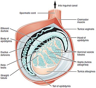
A testicle or testis is the male gonad in all bilaterians, including humans. It is homologous to the female ovary. The functions of the testicles are to produce both sperm and androgens, primarily testosterone. Testosterone release is controlled by the anterior pituitary luteinizing hormone, whereas sperm production is controlled both by the anterior pituitary follicle-stimulating hormone and gonadal testosterone.

<span class="mw-page-title-main">Cryptorchidism</span> Medical condition

Cryptorchidism, also known as undescended testis, is the failure of one or both testes to descend into the scrotum. The word is from Greek κρυπτός 'hidden' and ὄρχις 'testicle'. It is the most common birth defect of the male genital tract. About 3% of full-term and 30% of premature infant boys are born with at least one undescended testis. However, about 80% of cryptorchid testes descend by the first year of life, making the true incidence of cryptorchidism around 1% overall. Cryptorchidism may develop after infancy, sometimes as late as young adulthood, but that is exceptional.

Delayed puberty is when a person lacks or has incomplete development of specific sexual characteristics past the usual age of onset of puberty. The person may have no physical or hormonal signs that puberty has begun. In the United States, girls are considered to have delayed puberty if they lack breast development by age 13 or have not started menstruating by age 15. Boys are considered to have delayed puberty if they lack enlargement of the testicles by age 14. Delayed puberty affects about 2% of adolescents.

Hypogonadism means diminished functional activity of the gonads—the testicles or the ovaries—that may result in diminished production of sex hormones. Low androgen levels are referred to as hypoandrogenism and low estrogen as hypoestrogenism. These are responsible for the observed signs and symptoms in both males and females.

Kallmann syndrome (KS) is a genetic disorder that prevents a person from starting or fully completing puberty. Kallmann syndrome is a form of a group of conditions termed hypogonadotropic hypogonadism. To distinguish it from other forms of hypogonadotropic hypogonadism, Kallmann syndrome has the additional symptom of a total lack of sense of smell (anosmia) or a reduced sense of smell. If left untreated, people will have poorly defined secondary sexual characteristics, show signs of hypogonadism, almost invariably are infertile and are at increased risk of developing osteoporosis. A range of other physical symptoms affecting the face, hands and skeletal system can also occur.

<span class="mw-page-title-main">Spermatocele</span> Medical condition

Spermatocele is a fluid-filled cyst that develops in the epididymis. The fluid is usually a clear or milky white color and may contain sperm. Spermatoceles are typically filled with spermatozoa and they can vary in size from several millimeters to many centimeters. Small spermatoceles are relatively common, occurring in an estimated 30 percent of males. They are generally not painful. However, some people may experience discomfort such as a dull pain in the scrotum from larger spermatoceles. They are not cancerous, nor do they cause an increased risk of testicular cancer. Additionally, unlike varicoceles, they do not reduce fertility.

<span class="mw-page-title-main">Varicocele</span> Medical condition

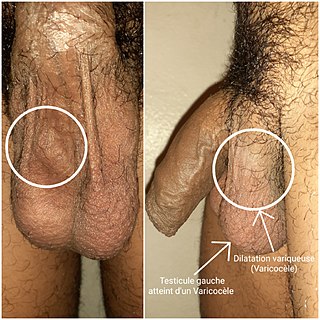
A varicocele is, in a male person, an abnormal enlargement of the pampiniform venous plexus in the scrotum; in a female person, it is an abnormal painful swelling to the embryologically identical pampiniform venous plexus; it is more commonly called pelvic compression syndrome. In the male varicocele, this plexus of veins drains blood from the testicles back to the heart. The vessels originate in the abdomen and course down through the inguinal canal as part of the spermatic cord on their way to the testis. Varicoceles occur in around 15% to 20% of all men. The incidence of varicocele increase with age.

<span class="mw-page-title-main">Azoospermia</span> Medical condition of a man whose semen contains no sperm

Azoospermia is the medical condition of a man whose semen contains no sperm. It is associated with male infertility, but many forms are amenable to medical treatment. In humans, azoospermia affects about 1% of the male population and may be seen in up to 20% of male infertility situations in Canada.

Terms oligospermia, oligozoospermia, and low sperm count refer to semen with a low concentration of sperm and is a common finding in male infertility. Often semen with a decreased sperm concentration may also show significant abnormalities in sperm morphology and motility. There has been interest in replacing the descriptive terms used in semen analysis with more quantitative information.

Hypospermia is a condition in which a man has an unusually low ejaculate volume, less than 1.5 mL. It is the opposite of hyperspermia, which is a semen volume of more than 5.5 mL. It should not be confused with oligospermia, which means low sperm count. Normal ejaculate when a man is not drained from prior sex and is suitably aroused is around 1.5–6 mL, although this varies greatly with mood, physical condition, and sexual activity. Of this, around 1% by volume is sperm cells. The U.S.-based National Institutes of Health defines hypospermia as a semen volume lower than 2 mL on at least two semen analyses.

Male infertility refers to a sexually mature male's inability to impregnate a fertile female. In humans, it accounts for 40–50% of infertility. It affects approximately 7% of all men. Male infertility is commonly due to deficiencies in the semen, and semen quality is used as a surrogate measure of male fecundity. More recently, advance sperm analyses that examine intracellular sperm components are being developed.

<span class="mw-page-title-main">Testicular sperm extraction</span> Surgical procedure

Testicular sperm extraction (TESE) is a surgical procedure in which a small portion of tissue is removed from the testicle and any viable sperm cells from that tissue are extracted for use in further procedures, most commonly intracytoplasmic sperm injection (ICSI) as part of in vitro fertilisation (IVF). TESE is often recommended to patients who cannot produce sperm by ejaculation due to azoospermia.

<span class="mw-page-title-main">Enclomifene</span> Chemical compound

Enclomifene (INNTooltip International Nonproprietary Name), or enclomiphene (USANTooltip United States Adopted Name), a nonsteroidal selective estrogen receptor modulator of the triphenylethylene group acts by antagonizing the estrogen receptor (ER) in the pituitary gland, which reduces negative feedback by estrogen on the hypothalamic-pituitary-gonadal axis, thereby increasing gonadotropin secretion and hence gonadal production of testosterone. It is one of the two stereoisomers of clomifene, which itself is a mixture of 38% zuclomifene and 62% enclomifene. Enclomifene is the (E)-stereoisomer of clomifene, while zuclomifene is the (Z)-stereoisomer. Whereas zuclomifene is more estrogenic, enclomifene is more antiestrogenic. In accordance, unlike enclomifene, zuclomifene is antigonadotropic due to activation of the ER and reduces testosterone levels in men. As such, isomerically pure enclomifene is more favorable than clomifene as a progonadotropin for the treatment of male hypogonadism.

<span class="mw-page-title-main">Orchiectomy</span> Surgical removal of one or both testicles

Orchiectomy is a surgical procedure in which one or both testicles are removed. The surgery can be performed for various reasons:

Hypergonadotropic hypogonadism (HH), also known as primary or peripheral/gonadal hypogonadism or primary gonadal failure, is a condition which is characterized by hypogonadism which is due to an impaired response of the gonads to the gonadotropins, follicle-stimulating hormone (FSH) and luteinizing hormone (LH), and in turn a lack of sex steroid production. As compensation and the lack of negative feedback, gonadotropin levels are elevated. Individuals with HH have an intact and functioning hypothalamus and pituitary glands so they are still able to produce FSH and LH. HH may present as either congenital or acquired, but the majority of cases are of the former nature. HH can be treated with hormone replacement therapy.

<span class="mw-page-title-main">Leydig cell hypoplasia</span> Medical condition

Leydig cell hypoplasia (LCH), also known as Leydig cell agenesis, is a rare autosomal recessive genetic and endocrine syndrome affecting an estimated 1 in 1,000,000 genetic males. It is characterized by an inability of the body to respond to luteinizing hormone (LH), a gonadotropin which is normally responsible for signaling Leydig cells of the testicles to produce testosterone and other androgen sex hormones. The condition manifests itself as pseudohermaphroditism, hypergonadotropic hypogonadism, reduced or absent puberty, and infertility.

Gonadotropin-releasing hormone (GnRH) insensitivity also known as Isolated gonadotropin-releasing hormone (GnRH)deficiency (IGD) is a rare autosomal recessive genetic and endocrine syndrome which is characterized by inactivating mutations of the gonadotropin-releasing hormone receptor (GnRHR) and thus an insensitivity of the receptor to gonadotropin-releasing hormone (GnRH), resulting in a partial or complete loss of the ability of the gonads to synthesize the sex hormones. The condition manifests itself as isolated hypogonadotropic hypogonadism (IHH), presenting with symptoms such as delayed, reduced, or absent puberty, low or complete lack of libido, and infertility, and is the predominant cause of IHH when it does not present alongside anosmia.

Follicle-stimulating hormone (FSH) insensitivity, or ovarian insensitivity to FSH in females, also referable to as ovarian follicle hypoplasia or granulosa cell hypoplasia in females, is a rare autosomal recessive genetic and endocrine syndrome affecting both females and males, with the former presenting with much greater severity of symptomatology. It is characterized by a resistance or complete insensitivity to the effects of follicle-stimulating hormone (FSH), a gonadotropin which is normally responsible for the stimulation of estrogen production by the ovaries in females and maintenance of fertility in both sexes. The condition manifests itself as hypergonadotropic hypogonadism, reduced or absent puberty, amenorrhea, and infertility in females, whereas males present merely with varying degrees of infertility and associated symptoms.

Androgen deficiency is a medical condition characterized by insufficient androgenic activity in the body. Androgen deficiency most commonly affects women, and is also called Female androgen insufficiency syndrome (FAIS), although it can happen in both sexes. Androgenic activity is mediated by androgens, and is dependent on various factors including androgen receptor abundance, sensitivity and function. Androgen deficiency is associated with lack of energy and motivation, depression, lack of desire (libido), and in more severe cases changes in secondary sex characteristics.

<span class="mw-page-title-main">Side effects of radiotherapy on fertility</span>

The side effects of radiotherapy on fertility are a growing concern to patients undergoing radiotherapy as cancer treatments. Radiotherapy is essential for certain cancer treatments and often is the first point of call for patients. Radiation can be divided into two categories: ionising radiation (IR) and non-ionising radiation (NIR). IR is more dangerous than NIR and a source of this radiation is X-rays used in medical procedures, for example in radiotherapy.

References

  1. 1 2 3 4 5 6 7 Ramos L, Patel AS, Ramasamy R (2018). "Testosterone replacement therapy for physician assistants and nurse practitioners". Translational Andrology and Urology. 7 (Suppl 1): S63–S71. doi: 10.21037/tau.2017.12.09 . PMC   5881199 . PMID   29644169.
  2. 1 2 3 4 5 Seal LJ (2013). "Male hypogonadism and testosterone replacement therapy". Medicine. 41 (10): 557–561. doi:10.1016/j.mpmed.2013.07.010 via Elsevier ScienceDirect.
  3. 1 2 3 4 Karagiannis A, Harsoulis F (2005). "Gonadal dysfunction in systemic diseases". European Journal of Endocrinology. 152 (4): 501–513. doi:10.1530/eje.1.01886. PMID   15817904. S2CID   27528976.
  4. 1 2 3 Rahnema CD, Lipshultz LI, Crosnoe LE, Kovac JR, Kim ED (2014). "Anabolic steroid-induced hypogonadism: diagnosis and treatment". Fertility and Sterility. 101 (5): 1271–1279. doi: 10.1016/j.fertnstert.2014.02.002 . PMID   24636400.
  5. 1 2 Cheng PJ, Pastuszak AW, Myers JB, Goodwin IA, Hotaling JM (2019). "Fertility concerns of the transgender patient". Translational Andrology and Urology. 8 (3): 209–218. doi: 10.21037/tau.2019.05.09 . PMC   6626312 . PMID   31380227.
  6. 1 2 3 4 DaJusta DG, Granberg CF, Villanueva C, Baker LA (2013). "Contemporary review of testicular torsion: new concepts, emerging technologies and potential therapeutics". Journal of Pediatric Urology. 9 (6 Pt A): 723–730. doi:10.1016/j.jpurol.2012.08.012. PMC   3566290 . PMID   23044376.
  7. 1 2 3 4 5 6 Garcia-Roig ML, Kirsch AJ (2015). "The dilemma of adolescent varicocele". Pediatric Surgery International. 31 (7): 617–625. doi:10.1007/s00383-015-3698-8. PMID   25895069. S2CID   2059903.
  8. 1 2 3 Selvaraj K, Ravichandran S, Krishnan S, Radhakrishnan RK, Manickam N, Kandasamy M (October 2021). "Testicular Atrophy and Hypothalamic Pathology in COVID-19: Possibility of the Incidence of Male Infertility and HPG Axis Abnormalities". Reproductive Sciences. 28 (10): 2735–2742. doi:10.1007/s43032-020-00441-x. PMC   7790483 . PMID   33415647.
  9. 1 2 3 Fijak M, Pilatz A, Hedger MP, Nicolas N, Bhushan S, Michel V, et al. (2018). "Infectious, inflammatory and 'autoimmune' male factor infertility: how do rodent models inform clinical practice?". Human Reproduction Update. 24 (4): 416–441. doi:10.1093/humupd/dmy009. PMC   6016649 . PMID   29648649.
  10. 1 2 Pedersen MR, Osther PJ, Rafaelsen SR (2018). "Ultrasound Evaluation of Testicular Volume in Patients with Testicular Microlithiasis". Ultrasound International Open. 4 (3): E99–E103. doi:10.1055/a-0643-4524. PMC   6143373 . PMID   30250943.
  11. 1 2 3 4 5 6 7 8 9 10 Fletcher J (2018). Murrell D (ed.). "Testicular atrophy: Causes, diagnosis, and treatment". Medical News Today. Retrieved 25 July 2022.
  12. 1 2 Misra M, Lee MM (2005). "Delayed puberty". Pediatric Endocrinology. Mosby. pp. 87–101. doi:10.1016/B978-0-323-01825-8.50035-1. ISBN   978-0-323-01825-8.
  13. Calabria A (2020). "Male Hypogonadism in Children - Pediatrics". Merck Manuals Professional Edition. Retrieved 1 August 2022.
  14. Hoffman M (2021). "Low Testosterone and Your Sex Drive". WebMD. Retrieved 2 August 2022.
  15. Griggs RC, Kingston W, Jozefowicz RF, Herr BE, Forbes G, Halliday D (January 1989). "Effect of testosterone on muscle mass and muscle protein synthesis". Journal of Applied Physiology. 66 (1): 498–503. doi:10.1152/jappl.1989.66.1.498. PMID   2917954.
  16. Medicover Hospitals (2021). "Testicular Atrophy Symptoms: Causes, Types, Treatments and Home Remedies". Medicover Hospitals. Retrieved 28 July 2022.
  17. 1 2 3 Moss JL, Crosnoe LE, Kim ED (2013). "Effect of rejuvenation hormones on spermatogenesis". Fertility and Sterility. 99 (7): 1814–1820. doi: 10.1016/j.fertnstert.2013.04.003 . PMID   23663992.
  18. Mieusset R, Bujan L, Plantavid M, Grandjean H (February 1989). "Increased levels of serum follicle-stimulating hormone and luteinizing hormone associated with intrinsic testicular hyperthermia in oligospermic infertile men". The Journal of Clinical Endocrinology and Metabolism. 68 (2): 419–425. doi: 10.1210/jcem-68-2-419 . PMID   2493031.
  19. 1 2 Macey MR, Owen RC, Ross SS, Coward RM (2018). "Best practice in the diagnosis and treatment of varicocele in children and adolescents". Therapeutic Advances in Urology. 10 (9): 273–282. doi:10.1177/1756287218783900. PMC   6088496 . PMID   30116303.
  20. "About Cystic Fibrosis | Cystic Fibrosis Foundation". Cystic Fibrosis Foundation. Retrieved 1 August 2022.
  21. Profka E, Rodari G, Giacchetti F, Berrettini A, Manzoni G, Daccò V, et al. (2020). "A case of testicular atrophy associated with cystic fibrosis". Endocrinology, Diabetes & Metabolism Case Reports. 2020: 20–0095. doi:10.1530/EDM-20-0095. PMC   7576656 . PMID   33434179.
  22. "Definition of vas deferens". National Cancer Institute. 2011. Retrieved 1 August 2022.
  23. Durell J, Johal N, Burge D, Wheeler R, Griffiths M, Kitteringham L, et al. (2016). "Testicular atrophy following paediatric primary orchidopexy: A prospective study". Journal of Pediatric Urology. 12 (4): 243.e1–243.e4. doi:10.1016/j.jpurol.2016.05.023. PMID   27422375.
  24. Winters SJ (2000). "Laboratory Assessment of Testicular Function". In Feingold KR, Anawalt B, Boyce A, Chrousos G (eds.). Endotext. South Dartmouth (MA): MDText.com, Inc. PMID   25905368 . Retrieved 26 July 2022.
  25. Kavoussi PK, Abdullah N, Gilkey MS, Hunn C, Machen GL, Chen SH, et al. (2021). "The impact of ipsilateral testicular atrophy on semen quality and sperm DNA fragmentation response to varicocele repair". Asian Journal of Andrology. 23 (2): 146–149. doi: 10.4103/aja.aja_50_20 . PMC   7991813 . PMID   32930104.
  26. Sharp VJ, Kieran K, Arlen AM (2013). "Testicular torsion: diagnosis, evaluation, and management". American Family Physician. 88 (12): 835–840. PMID   24364548.
  27. Aggarwal D, Parmar K, Sharma AP, Tyagi S, Kumar S, Singh SK, Gupta S (2022). "Long-term impact of testicular torsion and its salvage on semen parameters and gonadal function". Indian Journal of Urology. 38 (2): 135–139. doi: 10.4103/iju.iju_328_21 . PMC   8992720 . PMID   35400863.
  28. Leisegang K, Dutta S (2021). "Do lifestyle practices impede male fertility?". Andrologia. 53 (1): e13595. doi: 10.1111/and.13595 . hdl: 10566/5440 . PMID   32330362. S2CID   216129511.
  29. Skoracka K, Eder P, Łykowska-Szuber L, Dobrowolska A, Krela-Kaźmierczak I (2020). "Diet and Nutritional Factors in Male (In)fertility-Underestimated Factors". Journal of Clinical Medicine. 9 (5): 1400. doi: 10.3390/jcm9051400 . PMC   7291266 . PMID   32397485.
  30. Olza J, Aranceta-Bartrina J, González-Gross M, Ortega RM, Serra-Majem L, Varela-Moreiras G, Gil Á (2017). "Reported Dietary Intake and Food Sources of Zinc, Selenium, and Vitamins A, E and C in the Spanish Population: Findings from the ANIBES Study". Nutrients. 9 (7): 697. doi: 10.3390/nu9070697 . PMC   5537812 . PMID   28684689.