Triangular fibrocartilage

Last updated
Triangular fibrocartilage complex
Anatomy TFCC.jpg
Anatomy of the triangular fibrocartilage complex (TFCC)
Details
Identifiers
Latin discus articularis articulationis radioulnaris distalis
MeSH D051478
Anatomical terminology

The triangular fibrocartilage complex (TFCC) is formed by the triangular fibrocartilage discus (TFC), the radioulnar ligaments (RULs) and the ulnocarpal ligaments (UCLs).

Contents

Structure

Vertical section through the articulations at the wrist, showing the synovial cavities. (Articular disc labeled at center right.) Gray336.png
Vertical section through the articulations at the wrist, showing the synovial cavities. (Articular disc labeled at center right.)
Anatomy of the triangular fibrocartilage complex (TFCC) Anatomy TFCC 2.jpg
Anatomy of the triangular fibrocartilage complex (TFCC)

Triangular fibrocartilage disc

The triangular fibrocartilage disc (TFC) is an articular discus that lies on the pole of the distal ulna. It has a triangular shape and a biconcave body; the periphery is thicker than its center. The central portion of the TFC is thin and consists of chondroid fibrocartilage; this type of tissue is often seen in structures that can bear compressive loads. This central area is often so thin that it is translucent and in some cases it is even absent. [1] The peripheral portion of the TFC is well vascularized, while the central portion has no blood supply.

This discus is attached by thick tissue to the base of the ulnar styloid and by thinner tissue to the edge of the radius just proximal to the radiocarpal articular surface. [1]

Radioulnar ligaments

The radioulnar ligaments (RULs) are the principal stabilizers of the distal radioulnar joint (DRUJ). There are two RULs: the palmar and dorsal radioulnar ligaments.[ citation needed ]

These ligaments arise from the distal radius medial border and insert on the ulna at two separate and distinct sites: the ulna styloid and the fovea (a groove that separates the ulnar styloid from the ulnar head). Each ligament consists of a superficial component and a deep component. The superficial components insert directly onto the ulna styloid. The deep components insert more anterior, into the fovea adjacent to the articular surface of the dome of the distal ulna.[ citation needed ]

The ligaments are composed of longitudinally oriented lamellar collagen to resist tensile loads and have a rich vascular supply to allow healing. [2] [3] [4]

Ulnocarpal ligaments

The ulnocarpal ligaments (UCLs) consist of the ulnolunate and the ulnotriquetral ligaments. They originate from the ulnar styloid and insert into the carpal bones of the wrist: the ulnolunate ligament inserts into the lunate bone and the ulnotriquetral ligament into the triquetrum bone. These ligaments prevent dorsal migration of the distal ulna. They are more taut during supination, because in supination ulnar styloid moves away from the carpal bones volar side. [1] [5] [6]

Function

The primary functions of the TFCC:

Load transmission

The TFCC is important in load transmission across the ulnar aspect of the wrist. The TFC transmits and absorbs compressive forces.

The ulnar variance influences the amount of load that is transmitted through the distal ulna. The load transmission is directly proportional to this ulnar variance. In neutral ulnar variance, approximately 20 percent of the load is transmitted. With negative ulnar variance, the load across the TFC is decreased. This occurs during supination, because the radius moves distally on the ulna and creates a negative ulnar variance. With positive ulnar variance it is reversed. The load that is transmitted across the TFC is then increased. This positive ulnar variance occurs during pronation. [7]

Rotation

Rul's prosupination Rul's by pro- and supination.jpg
Rul's prosupination

The TFCC is a major stabilizer of the DRUJ. To control the forearm rotation the DRUJ acts in concert with the proximal radioulnar joint. The connection between the distal radius and the distal ulna, maintain the congruency of the DRUJ. This attachment is mainly created by the RULs of the TFCC. These ligaments support the joint through its arc of rotation. [7]

The role of the TFCC in supination and in pronation is a matter of dispute. Some authors (Schuind et al.) concluded that the dorsal fibers of the TFCC tighten in pronation, and the palmar fibers in supination. These conclusions are opposite of those published by Af Ekenstam and Hagert. [2]

Both parties are in fact right, as the RULs consists of two ligaments each made of another two components: the superficial and the deep ligaments. During supination, the superficial palmar and the deep dorsal ligaments are tightened,[ citation needed ] preventing palmar translation of the ulna. In pronation, this is reversed: the superficial dorsal and the deep palmar ligaments are tightened and prevent dorsal translation of the ulna.[ citation needed ]

Clinical significance

The TFCC has a substantial risk for injury and degeneration because of its anatomic complexity and multiple functions.

Application of an extension-pronation force to an axial-load wrist, such as in a fall on an outstretched hand, causes most of the traumatic injuries of the TFCC.

Dorsal rotation injury, such as when a drill binds and rotates the wrist instead of the bit, can also cause traumatic injuries.

Injury may also occur from a distraction force applied to the volar forearm or wrist. Finally, tears of the TFCC are frequently found by patients with distal radius fractures. [7]

Perforations and defects in the TFCC are not all traumatic. There is an age related correlation with lesions in the TFCC, but many of these defects are asymptomatic. These lesions common occur by patients with positive ulnar variance. [7]

Chronic and excessive loading through the ulnocarpal joint, causes degenerative TFCC tears. These tears are a component of ulnar impaction syndrome.

Even though natural degeneration of the ulnocarpal joint is very common, it is important to recognize. In cadavaric examinations, 30% to 70% of the cases had TFCC perforations and chondromalacia of the ulnar head, lunate, and triquetrum. Cases with ulnar-negative variance had fewer degenerative changes. [1]

Palmer classification of TFCC lesions

The Palmer classification is the most recognized scheme; it divides TFCC lesions into these two categories: traumatic and degenerative. This classification provides an anatomic description of tears, it does not guide treatment or indicate prognosis. [1]

Class 1 – Traumatic
Class 1A. Central perforation
Class 1B. Ulnar avulsion (with or without styloid fracture)
Class 1C. Distal avulsion (from carpus)
Class 1D. Radial avulsion (with or without sigmoid notch fracture)
Class 2 – Degenerative (ulnar impaction syndrome)
Class 2A. TFCC wear
Class 2B. TFCC wear with lunate and/or ulnar head chondromalacia
Class 2C. TFCC perforation with lunate and/or ulnar head chondromalacia
Class 2D. TFCC perforation with lunate and/or ulnar head chondromalacia, and with lunotriquetral ligament perforation
Class 2E. TFCC perforation with lunate and/or ulnar head chondromalacia, with lunotriquetral ligament perforation, and with ulnocarpal arthritis

Symptoms

Patients with a TFCC injury usually experience pain or discomfort located at the ulnar side of the wrist, often just above the ulnar styloid. However, there are also some patients who report diffuse pain throughout the entire wrist.

Rest can reduce pain and activity can make it worse, especially with rotating movements (supination and pronation) of the wrist or movements of the hand sideways in ulnar direction.

Other symptoms patients with a TFCC injury frequently mention are: swelling, loss of grip strength, instability, and grinding or clicking sounds (crepitus) that can occur during activity of the wrist. [8]

Diagnosis

Anamnesis

Injuries to the TFCC may be preceded by a fall on a pronated outstretched arm; a rotational injury to the forearm; an axial load trauma to the wrist; or a distraction injury of the wrist in ulnar direction. [8] However, not all patients can recall that a preceding trauma occurred.

Physical examination

Imaging

Arthroscopic image of a central triangular fibrocartilage complex (TFCC) tear TFCC tear.jpg
Arthroscopic image of a central triangular fibrocartilage complex (TFCC) tear
  • X-ray: X-rays of the wrist are made in two directions: posterior-anterior (PA) and lateral. Radiographs are useful to diagnose or rule out possible bone fractures, a positive ulnar variance or osteoarthritis. The TFCC is not visible on an X-ray, regardless of its condition.
  • MRI: is, together with the findings of a careful physical examination, a helpful diagnostic tool to assess the condition of the TFCC. Nevertheless, the incidence of false-positive and false-negative MRI results is high. [2]
  • Arthrography: a dye is injected into the wrist joint. If there is a TFCC lesion the dye will leak from one joint compartment to another.
  • Wrist arthroscopy: is an invasive diagnostic tool, but it remains to this day the most accurate way to identify TFCC lesions. [9]

Note: Imaging techniques can only be relevant together with the clinical findings of a carefully performed physical examination.

Other than a TFCC injury, there are many possible causes for ulnar-sided wrist pain.

Differential diagnosis of TFCC injuries

  • Tendinopathy of the ECU
  • Ulnar styloid fracture
  • Distal radius fracture
  • DRUJ arthritis
  • Pisiform bone fractures
  • Hamate bone fractures
  • Carpal instability
  • Midcarpal instability
  • Hypothenar hammer syndrome (ulnar artery thrombosis)

Treatment

The initial treatment for both traumatic and degenerative TFCC lesions, with a stable DRUJ, is conservative (nonsurgical) therapy. Patients may be advised to wear a temporary splint or cast to immobilize the wrist and forearm for four to six weeks. [10] The immobilization allows scar tissue to develop which can help heal the TFCC. In addition, oral NSAIDs and corticosteroid joint injections can be prescribed for pain relief. Physiotherapy and occupational therapy can help patients recover after immobilization or surgery. Wrist support straps used in sports can also be used in mild cases to compress and minimize movement of the area. [11]

Indications for acute TFCC surgery are: a clearly unstable DRUJ, or the existence of additional unstable or displaced fractures. TFCC surgery is also indicated when conservative treatment proves insufficient in about 8–12 weeks.

Fractures of the radius bone are often associated by TFCC damage. If the fracture is treated surgically it is recommended to evaluate and if necessary repair the TFCC as well. [12] Closed fractures (where the skin is still intact) of the radius bone are treated non-surgically with cast; the immobilization can also help heal the TFCC.

Surgical

Arthroscopic debridement of TFC discus tissue

The central part of the TFC has no blood supply and therefore has no healing capacity. When a tear occurs in this area of the TFC, it typically creates an unstable flap of tissue that is likely to catch on other joint surfaces. Removing the damaged tissue (debridement) is then indicated. Arthroscopic debridement as a treatment for degenerative TFC tears associated with positive ulnar variance, unfortunately, show poor results. [10] [9]

Arthroscopic repair of TFCC ligaments

Suturing TFCC ligaments can sometimes be performed arthroscopically. But only if there is no serious damage to the ligaments or other surrounding structures. Even after a short period of time torn ligaments tend to retract and therefore lose length. Retracted ligament ends are impossible to suture together again and a reconstruction may be necessary.[ citation needed ]

Open surgical repair of the TFCC

Open surgery is usually required for degenerative or more complex TFCC injuries, or if additional damage to the wrist or forearm caused instability or displacement. It is a more invasive surgical technique compared to arthroscopic treatment, but the surgeon has better visibility and access to the TFCC.[ citation needed ]

Options for open surgery
  • Suturing of the RULs. This is, just like arthroscopic suturing of these ligaments, only possible when the damage is not too serious and if both ends of the ruptured ligament are not yet retracted.
  • Anatomic reconstruction of the RULs using a tendon graft (e.g., the palmaris longus). The tendon graft is tunneled through drilled holes in the ulnar and radius bones. This procedure is indicated for DRUJ instability caused by an irreparable TFCC. [13] [3]
  • Capsular or extensor retinaculum plication. This surgical technique aims to improve DRUJ stability by shortening the joint capsule or the extensor retinaculum. It is mostly used for minor DRUJ instability and is less invasive compared to a complete RUL reconstruction. [14] [15]
  • Shortening of the ulnar bone. Patients with a positive ulnar variance are more susceptible to TFCC damage. Shortening the ulnar bone may help relieve the excess pressure to the TFCC and prevent further degeneration. [16]

Related Research Articles

<span class="mw-page-title-main">Carpal bones</span> Eight small bones that make up the wrist (or carpus) that connects the hand to the forearm

The carpal bones are the eight small bones that make up the wrist (carpus) that connects the hand to the forearm. The term "carpus" and "carpal" is derived from the Latin carpus and the Greek καρπός (karpós), meaning "wrist". In human anatomy, the main role of the carpal bones is to articulate with the radial and ulnar heads to form a highly mobile condyloid joint, to provide attachments for thenar and hypothenar muscles, and to form part of the rigid carpal tunnel which allows the median nerve and tendons of the anterior forearm muscles to be transmitted to the hand and fingers.

<span class="mw-page-title-main">Ulna</span> Medial bone from forearm

The ulna or ulnar bone is a long bone in the forearm stretching from the elbow to the wrist. It is on the same side of the forearm as the little finger, running parallel to the radius, the forearm's other long bone. Longer and thinner than the radius, the ulna is considered to be the smaller long bone of the lower arm. The corresponding bone in the lower leg is the fibula.

<span class="mw-page-title-main">Wrist</span> Part of the arm between the lower arm and the hand

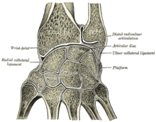
In human anatomy, the wrist is variously defined as (1) the carpus or carpal bones, the complex of eight bones forming the proximal skeletal segment of the hand; (2) the wrist joint or radiocarpal joint, the joint between the radius and the carpus and; (3) the anatomical region surrounding the carpus including the distal parts of the bones of the forearm and the proximal parts of the metacarpus or five metacarpal bones and the series of joints between these bones, thus referred to as wrist joints. This region also includes the carpal tunnel, the anatomical snuff box, bracelet lines, the flexor retinaculum, and the extensor retinaculum.

The forearm is the region of the upper limb between the elbow and the wrist. The term forearm is used in anatomy to distinguish it from the arm, a word which is used to describe the entire appendage of the upper limb, but which in anatomy, technically, means only the region of the upper arm, whereas the lower "arm" is called the forearm. It is homologous to the region of the leg that lies between the knee and the ankle joints, the crus.

<span class="mw-page-title-main">Scaphoid bone</span> Bone of the carpus

The scaphoid bone is one of the carpal bones of the wrist. It is situated between the hand and forearm on the thumb side of the wrist. It forms the radial border of the carpal tunnel. The scaphoid bone is the largest bone of the proximal row of wrist bones, its long axis being from above downward, lateralward, and forward. It is approximately the size and shape of a medium cashew nut.

<span class="mw-page-title-main">Radius (bone)</span> One of the two long bones of the forearm

The radius or radial bone is one of the two large bones of the forearm, the other being the ulna. It extends from the lateral side of the elbow to the thumb side of the wrist and runs parallel to the ulna. The ulna is longer than the radius, but the radius is thicker. The radius is a long bone, prism-shaped and slightly curved longitudinally.

<span class="mw-page-title-main">Triquetral bone</span> Bone in the wrist

The triquetral bone is located in the wrist on the medial side of the proximal row of the carpus between the lunate and pisiform bones. It is on the ulnar side of the hand, but does not directly articulate with the ulna. Instead, it is connected to and articulates with the ulna through the Triangular fibrocartilage disc and ligament, which forms part of the ulnocarpal joint capsule. It connects with the pisiform, hamate, and lunate bones. It is the 2nd most commonly fractured carpal bone.

<span class="mw-page-title-main">Distal radius fracture</span> Fracture of the radius bone near the wrist

A distal radius fracture, also known as wrist fracture, is a break of the part of the radius bone which is close to the wrist. Symptoms include pain, bruising, and rapid-onset swelling. The ulna bone may also be broken.

<span class="mw-page-title-main">Galeazzi fracture</span> Medical condition

The Galeazzi fracture is a fracture of the distal third of the radius with dislocation of the distal radioulnar joint. It classically involves an isolated fracture of the junction of the distal third and middle third of the radius with associated subluxation or dislocation of the distal radio-ulnar joint; the injury disrupts the forearm axis joint.

<span class="mw-page-title-main">Madelung's deformity</span> Medical condition

Madelung's deformity is usually characterized by malformed wrists and wrist bones and is often associated with Léri-Weill dyschondrosteosis. It can be bilateral or just in the one wrist. It has only been recognized within the past hundred years. Named after Otto Wilhelm Madelung (1846–1926), a German surgeon, who described it in detail, it was noted by others. Guillaume Dupuytren mentioned it in 1834, Auguste Nélaton in 1847, and Joseph-François Malgaigne in 1855.

<span class="mw-page-title-main">Annular ligament of radius</span>

The annular ligament is a strong band of fibers that encircles the head of the radius, and retains it in contact with the radial notch of the ulna.

<span class="mw-page-title-main">Distal radioulnar articulation</span>

The distal radioulnar articulation is a synovial pivot joint between the two bones in the forearm; the radius and ulna. It is one of two joints between the radius and ulna, the other being the proximal radioulnar articulation. The joint features an articular disc, and is reinforced by the palmar and dorsal radioulnar ligaments.

<span class="mw-page-title-main">Ulnar styloid process</span> Bony prominence at the wrist

The styloid process of the ulna is a bony prominence found at distal end of the ulna in the forearm.

<span class="mw-page-title-main">Interosseous membrane of forearm</span>

The interosseous membrane of the forearm is a fibrous sheet that connects the interosseous margins of the radius and the ulna. It is the main part of the radio-ulnar syndesmosis, a fibrous joint between the two bones.

<span class="mw-page-title-main">Midcarpal joint</span>

The midcarpal joint is formed by the scaphoid, lunate, and triquetral bones in the proximal row, and the trapezium, trapezoid, capitate, and hamate bones in the distal row. The distal pole of the scaphoid articulates with two trapezial bones as a gliding type of joint. The proximal end of the scaphoid combines with the lunate and triquetrum to form a deep concavity that articulates with the convexity of the combined capitate and hamate in a form of diarthrodial, almost condyloid joint.

<span class="mw-page-title-main">Elbow</span> Joint between the upper and lower parts of the arm

The elbow is the region between the upper arm and the forearm that surrounds the elbow joint. The elbow includes prominent landmarks such as the olecranon, the cubital fossa, and the lateral and the medial epicondyles of the humerus. The elbow joint is a hinge joint between the arm and the forearm; more specifically between the humerus in the upper arm and the radius and ulna in the forearm which allows the forearm and hand to be moved towards and away from the body. The term elbow is specifically used for humans and other primates, and in other vertebrates it is not used. In those cases, forelimb plus joint is used.

The Essex-Lopresti fracture is a fracture of the radial head of the forearm with concomitant dislocation of the distal radio-ulnar joint along with disruption of the thin interosseous membrane which holds them together. The injury is named after Peter Essex-Lopresti who described it in 1951.

<span class="mw-page-title-main">Wrist osteoarthritis</span> Medical condition

Wrist osteoarthritis is gradual loss of articular cartilage and hypertrophic bone changes (osteophytes). While in many joints this is part of normal aging (senescence), in the wrist osteoarthritis usually occurs over years to decades after scapholunate interosseous ligament rupture or an unhealed fracture of the scaphoid. Characteristic symptoms including pain, deformity and stiffness. Pain intensity and incapability are notably variable and do not correspond with arthritis severity on radiographs.

<span class="mw-page-title-main">Wrist arthroscopy</span>

Wrist arthroscopy can be used to look inside the joint of the wrist. It is a minimally invasive technique which can be utilized for diagnostic purposes as well as for therapeutic interventions. Wrist arthroscopy has been used for diagnostic purposes since it was first introduced in 1979. However, it only became accepted as diagnostic tool around the mid-1980s. At that time, arthroscopy of the wrist was an innovative technique to determine whether a problem could be found in the wrist. A few years later, wrist arthroscopy could also be used as a therapeutic tool.

<span class="mw-page-title-main">Classification of distal radius fractures</span>

There are a number of ways to classify distal radius fractures. Classifications systems are devised to describe patterns of injury which will behave in predictable ways, to distinguish between conditions which have different outcomes or which need different treatments. Most wrist fracture systems have failed to accomplish any of these goals and there is no consensus about the most useful one.

References

PD-icon.svgThis article incorporates text in the public domain from page 325 of the 20th edition of Gray's Anatomy (1918)

  1. 1 2 3 4 5 Adams, Brian D.; Leversedge, Fraser J. (2016). "Distal Radioulnar Joint". In Wolfe, Scott W.; Pederson, William C.; Hotchkiss, Robert N.; Kozin, Scott H.; Cohen, Mark S (eds.). Green's Operative Hand Surgery (7th ed.). pp. 479–515. ISBN   978-0-323-29534-5.
  2. 1 2 3 4 Kleinman, William B (2007). "Stability of the Distal Radioulna Joint: Biomechanics, Pathophysiology, Physical Diagnosis, and Restoration of Function What We Have Learned in 25 Years". The Journal of Hand Surgery. 32 (7): 1086–106. doi:10.1016/j.jhsa.2007.06.014. PMID   17826566.
  3. 1 2 Lawler, Ericka; Adams, Brian D (2007). "Reconstruction for DRUJ Instability". HAND. 2 (3): 123–6. doi:10.1007/s11552-007-9034-6. PMC   2527144 . PMID   18780072.
  4. Gutiérrez, Alejandro Espinosa; Montero, José A. Rivas; Gutiérrez, Luis Enrique Sánchez (2007). "Inestabilidad radiocubital distal: Reconstrucción del ligamento: dorsal del fibrocartílago triangular y su evolución" [Instability from the distal radioulnar joint: Reconstruction and evolution of dorsal triangular fibrocartilage complex ligament]. Acta Ortopedica Mexicana (in Spanish). 21 (5): 282–8. PMID   18159918.
  5. Ishii, Shoji; Palmer, Andrew K; Werner, Frederick W; Short, Walter H; Fortino, Maria D (1998). "An anatomic study of the ligamentous structure of the triangular fibrocartilage complex". The Journal of Hand Surgery. 23 (6): 977–85. doi:10.1016/S0363-5023(98)80003-8. PMID   9848546.
  6. Manaster, B. J., Julia Crim "Imaging Anatomy: Musculoskeletal E-Book" Elsevier Health Sciences, 2016, p. 326.
  7. 1 2 3 4 Geissler, William B. (2016). "Wrist Arthroscopy". In Wolfe, Scott W.; Pederson, William C.; Hotchkiss, Robert N.; Kozin, Scott H.; Cohen, Mark S (eds.). Green's Operative Hand Surgery (7th ed.). pp. 653–96. ISBN   978-0-323-29534-5.
  8. 1 2 3 Ahn, A. K; Chang, D; Plate, A. M (2006). "Triangular fibrocartilage complex tears: A review" (PDF). Bulletin of the NYU Hospital for Joint Diseases. 64 (3–4): 114–8. PMID   17155920.
  9. 1 2 Elkowitz, S. J; Posner, M. A (2006). "Wrist arthroscopy" (PDF). Bulletin of the NYU Hospital for Joint Diseases. 64 (3–4): 156–65. PMID   17155924.
  10. 1 2 Henry, M. H (2008). "Management of acute triangular fibrocartilage complex injury of the wrist". The Journal of the American Academy of Orthopaedic Surgeons. 16 (6): 320–9. doi:10.5435/00124635-200806000-00004. PMID   18524983. S2CID   28702995.
  11. Barlow, Susan J (2016). "A Non-surgical Intervention for Triangular Fibrocartilage Complex Tears". Physiotherapy Research International. 21 (4): 271–276. doi:10.1002/pri.1672. PMID   27196674.
  12. Argintar, Evan; Mantovani, Gustavo; Pavan, Alexandre (2010). "TFCC Reattachment After Traumatic DRUJ Instability". Techniques in Hand & Upper Extremity Surgery. 14 (4): 226–9. doi:10.1097/BTH.0b013e3181e34fee. PMID   21107219. S2CID   32848551.
  13. Adams, Brian D; Berger, Richard A (2002). "An anatomic reconstruction of the distal radioulnar ligaments for posttraumatic distal radioulnar joint instability". The Journal of Hand Surgery. 27 (2): 243–51. doi:10.1053/jhsu.2002.31731. PMID   11901383.
  14. Johnston, K; Durand, D; Hildebrand, K. A (2009). "Chronic volar distal radioulnar joint instability: Joint capsular plication to restore function". Canadian Journal of Surgery. 52 (2): 112–8. PMC   2663503 . PMID   19399205.
  15. Wong, Kwok-Ho; Yip, Tak-Hing; Wu, Wing-Cheung (2004). "Distal Radioulnar Joint Dorsal Instability Treated with Dorsal Capsular Reconstruction". Hand Surgery. 9 (1): 55–61. doi:10.1142/S0218810404002133. PMID   15368627.
  16. Jui-Tien, Shih; Yao-Tung, Hou; Hung-Maan, Lee; Chuan-Ming, Tan; Ming-Chun, Chang (2016). "Chronic Triangular Fibrocartilage Complex Tears with Distal Radioulna Joint Instability: A New Method of Triangular Fibrocartilage Complex Reconstruction". Journal of Orthopaedic Surgery. 8 (1): 1–8. doi: 10.1177/230949900000800102 . PMID   12468868.