Trichoderma koningii

Last updated

Trichoderma koningii
Archives neerlandaises des sciences exactes et naturelles (1902) (19704647794).jpg
Scientific classification OOjs UI icon edit-ltr.svg
Domain: Eukaryota
Kingdom: Fungi
Division: Ascomycota
Class: Sordariomycetes
Order: Hypocreales
Family: Hypocreaceae
Genus: Trichoderma
Species:
T. koningii
Binomial name
Trichoderma koningii
Oudem. (1902)
Synonyms
  • Acrostalagmus koningii(Oudem.) Duché & R. Heim (1931)

Trichoderma koningii is a very common soil dwelling saprotroph with a worldwide distribution. [1] It has been heavily exploited for agricultural use as an effective biopesticide, having been frequently cited as an alternative biological control agent in the regulation of fungi-induced plant diseases. [2] [3] They are endosymbionts associated with plant root tissues, exhibiting mycoparasitism and promoting plant growth due to their capacity to produce different secondary metabolites. [4]

Contents

Trichoderma koningii is a species belonging to the genus Trichoderma . Fungi in this genus are able to adapt to different ecological niches and can colonize their habitats effectively, allowing them to be powerful antagonists and biocontrol agents. [5] Typical of Trichoderma species is having a fast growth rate and the production of green or hyaline conidia on a branched conidiophore structure. [5] [6]

History and taxonomy

Trichoderma koningii was first described by the Dutch mycologist Oudemans in 1902 as one of the species in the microbial flora he obtained from a nature preserve in The Netherlands. [7] After the genus was erected in 1794, there was difficulty in distinguishing and identifying the different species apart due to their very similar morphological characteristics. It wasn't until 1969 that a concept of classification was proposed by Rifai to reduce confusion on the taxonomy of Trichoderma. [5] He recognized T. koningii as one of the nine "aggregates" or groups of species in the genus. This aggregate consists of 12 species within three lineages that have similar morphology as the "true" T. koningii but can be differentiated from each other by their phenotypic characters and geographic distributions. [3] In 1991, Bissett divided the genus into five sections to classify the species on the basis of the branching of conidiophores. He included T. koningii in Trichoderma sect. Trichoderma. [8] In 2004, Chaverri and Samuels proposed another taxonomic classification based on molecular phylogenetic analysis. T. koningii and its aggregates were included in the T. viride clade. [3]

Growth and morphology

Brown, wrinkled stromata seen in some Hypocrea species. Hypocrea sp. (44271230445).jpg
Brown, wrinkled stromata seen in some Hypocrea species.

The conidiophores of T. koningii are branched and organized in a pyramidal structure with longer branches at the base that progressively shortens as it nears the tip. Primary and secondary branches arise in a right-angle degree and are often symmetrical on either side of the node along the main axis. Phialides are usually in 3–4 whorls that arise from the tip of the main branch and from lateral branches at intercalary positions on the conidiophore. [1] Some phialides on widely-spaced branches are flask-shaped, resembling a wine bottle, whereas some tend to have a very swollen middle when in dense clusters or "pseudo-whorls". [3] T. koningii typically produces smooth and ellipsoidal (egg-shaped) conidia, with a mean length of 4.1–4.3 µm, that aggregates in a slimy green mass at the tip of the phialides. The chlamydospores are pale brown, globe-like in shape, and are located at terminal and intercalary positions on the hyphae. [9]

In culture, colonies display rapid growth on potato dextrose agar (PDA), as cream-coloured in the beginning but later turns green because of sporulation. [10] [9] T. koningii grows at an optimum temperature of 25 °C in darkness, producing white mycelium with a radius of 50–60 mm. During conidial production, colouration first begins at the centre then later spreads outward in dark or dull green concentric rings that are vague to noticeable. [3] Maximum temperature for growth is observed at 33 °C, which reduces their pathogenic potential in humans. [11]

Like most Trichoderma species, this fungus has a sexual state. The teleomorph, Hypocrea koningii, is characterized by cushion-shaped stromata (sing. stroma) that are broadly attached to the surface of a substrate but are free at the margins. The surface of the stroma appears slightly-wrinkled. Mature stromata are brown to brownish-orange, whereas the young ones have a tan color with villi sprouting from the surface. These short hairs are lost during development. Perithecia (fruiting bodies) are elliptic, 160–280 µm long and 100–185 µm wide. The perithecial neck has a length of 53–90 µm. Asci (sing. ascus) within the fruiting bodies are typically cylindrical, with dimensions of 60–70 x 4–5.7 µm and thickening at the apex. The ascospores of H. koningii are hyaline and fill up the ascus in a single row. They are initially bicellular but have become separated into part-ascospores. The proximal part of the ascospore is ellipsoidal while the distal part is globe-like and longer. [1] [3]

Physiology

Trichoderma koningii is employed as a biological control agent because of its mycoparasitic and antagonistic ability. This fungus is capable of biosynthesizing silver nanoparticles, volatile organic compounds and secondary metabolites such as trichokonins, koninginins, and pyrones. [5] Silver nanoparticles (AgNPs) are produced via the reduction and capping of Ag+ to Ag0 by the enzymes and proteins released by T. koningii. [12] Koninginins are substances capable of inhibiting the process of inflammation. Koninginins isolated from T. koningii are identified to be A, E, F, L and M (KonA, KonE, KonF, KonL, KonM). [13] Trichokonins are peptaibols that exhibit antimicrobial property. Other polyketides reportedly isolated from T. koningii are Trichodermaketones A-D, 7-O-Methylkoninginin D, and 6-pentyl alpha pyrone which can inhibit the germination of other fungal spores. [3] [14] [15]

T. koningii is also reported to produce calcium oxalate crystals, particularly weddellite, via biomineralization. The process occurs intracellularly and extracellularly with respect to the fungus. The intracellular process involves the vegetative growth of the mycelium. The extracellular activity occurs through the reaction between the calcium in the environment and oxalic acid secreted by the fungus, leading to the production of biomineral species. [16]

Habitat and ecology

Typical of Trichoderma, T. koningii is a good colonizer of its habitat. Saprophytic growth occurs in acidified soils and soils with high water content (i.e. chernozem, podzol). [17] It is often isolated from under pine and coniferous trees, vegetation, plantations, grasslands, marshes, swamps, and peats. T. koningii also thrives in other environments, including growing on decaying wood, in marine species, estuarine sediments, and in mines and caves. [6] [10] The fruiting bodies commonly grow on tree bark [3] and stromata tend to be scattered, often solitary than in clusters. [1] It is distributed widely in Europe, the United States, and Canada. [3] Recent surveys have reported that some strains of T. koningii are also found to be present in New Zealand [4] and South Africa. [6]

Applications

Agriculture

Trichoderma koningii are plant symbionts that induce resistance against fungal pathogen attack and stimulate growth. It acts as a parasite to other fungi, particularly those that cause diseases to plants, by inhibiting their growth or attacking them directly. It is antagonistic to various plant pathogens such as Gaeumannomyces graminis var. tritici (Ggt), Sclerotium rolfsii , and Sclerotium cepivorum . It inhibits the growth of Ggt by releasing its microbial compounds. It colonizes the rhizospheres to interact with the roots of seedlings and plants, preventing S. rolfsii from damping-off the seedlings before they can germinate. [18] T. koningii antagonizes S. cepivorum by acting as a secondary colonizer of the infected plant roots and secreting enzymes that cause the degradation and lysis of the pathogen. [19] [20]

Medicine

Several studies have described the ability of T. koningii to produce enzymes that exhibit antifungal and antibacterial properties. Koninginins bear similar structural elements as flavonoids and vitamin E. They can inhibit the process of inflammation caused by snake bites. They can block the effects of myotoxins and induction of edema because they can inhibit phospholipase A2, one of the proteins found in venoms. [13] AgNPs produced using T. koningii are recognized as alternatives to antibiotics and are tools for gene delivery and drug delivery. They also show antagonism against Gram-positive and Gram-negative bacteria, respectively Candida albicans and Salmonella typhimurium . [12]

Related Research Articles

<span class="mw-page-title-main">Sclerotium</span> Mycelial mass

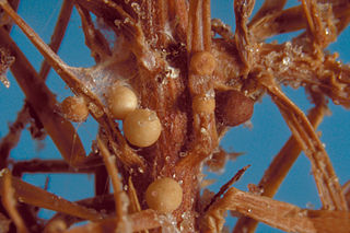
A sclerotium, is a compact mass of hardened fungal mycelium containing food reserves. One role of sclerotia is to survive environmental extremes. In some higher fungi such as ergot, sclerotia become detached and remain dormant until favorable growth conditions return. Sclerotia initially were mistaken for individual organisms and described as separate species until Louis René Tulasne proved in 1853 that sclerotia are only a stage in the life cycle of some fungi. Further investigation showed that this stage appears in many fungi belonging to many diverse groups. Sclerotia are important in the understanding of the life cycle and reproduction of fungi, as a food source, as medicine, and in agricultural blight management.

Tolypocladium inflatum is an ascomycete fungus originally isolated from a Norwegian soil sample that, under certain conditions, produces the immunosuppressant drug ciclosporin. In its sexual stage (teleomorph) it is a parasite on scarab beetles. It forms a small, compound ascocarp that arises from the cadaver of its host beetle. In its asexual stage (anamorph) it is a white mold that grows on soil. It is much more commonly found in its asexual stage and this is the stage that was originally given the name Tolypocladium inflatum.

<i>Trichoderma</i> Genus of fungi

Trichoderma is a genus of fungi in the family Hypocreaceae that is present in all soils, where they are the most prevalent culturable fungi. Many species in this genus can be characterized as opportunistic avirulent plant symbionts. This refers to the ability of several Trichoderma species to form mutualistic endophytic relationships with several plant species. The genomes of several Trichoderma specieshave been sequenced and are publicly available from the JGI.

<i>Epichloë</i> Genus of fungi

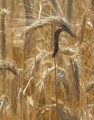
Epichloë is a genus of ascomycete fungi forming an endophytic symbiosis with grasses. Grass choke disease is a symptom in grasses induced by some Epichloë species, which form spore-bearing mats (stromata) on tillers and suppress the development of their host plant's inflorescence. For most of their life cycle however, Epichloë grow in the intercellular space of stems, leaves, inflorescences, and seeds of the grass plant without incurring symptoms of disease. In fact, they provide several benefits to their host, including the production of different herbivore-deterring alkaloids, increased stress resistance, and growth promotion.

<i>Acrophialophora fusispora</i> Species of ascomycete fungus found in soil, air and various plants

Acrophialophora fusispora is a poorly studied ascomycete fungus found in soil, air and various plants. A. fusispora is morphologically similar to the genera Paecilomyces and Masonia, but differ in the presence of pigmented conidiophores, verticillate phialides, and frequent sympodial proliferation. Moreover, A. fusispora is distinguished by its pigmented spindle-shaped conidia, covered with spiral bands. The fungus is naturally found in soils of tropical to temperate regions. The fungus has been identified as a plant and animal pathogen, and has recently been recognized as an emerging opportunistic human pathogen. A. fusispora infection in human is rare and has few documented clinical cases, but due to the rarity of the fungus and potential misidentification, the infections may be underdiagnosed. Clinical cases of A. fusispora include cases of keratitis, pulmonary colonization and infection, and cerebral infections. The fungus also has two documented cases of infection in dogs.

<i>Geotrichum candidum</i> Species of fungus

Geotrichum candidum is a fungus which is a member of the human microbiome, notably associated with skin, sputum, and faeces where it occurs in 25–30% of specimens. It is common in soil and has been isolated from soil collected around the world, in all continents.

Aspergillus ochraceus is a mold species in the genus Aspergillus known to produce the toxin ochratoxin A, one of the most abundant food-contaminating mycotoxins, and citrinin. It also produces the dihydroisocoumarin mellein. It is a filamentous fungus in nature and has characteristic biseriate conidiophores. Traditionally a soil fungus, has now began to adapt to varied ecological niches, like agricultural commodities, farmed animal and marine species. In humans and animals the consumption of this fungus produces chronic neurotoxic, immunosuppressive, genotoxic, carcinogenic and teratogenic effects. Its airborne spores are one of the potential causes of asthma in children and lung diseases in humans. The pig and chicken populations in the farms are the most affected by this fungus and its mycotoxins. Certain fungicides like mancozeb, copper oxychloride, and sulfur have inhibitory effects on the growth of this fungus and its mycotoxin producing capacities.

Coniochaeta hoffmannii, also known as Lecythophora hoffmannii, is an ascomycete fungus that grows commonly in soil. It has also been categorized as a soft-rot fungus capable of bringing the surface layer of timber into a state of decay, even when safeguarded with preservatives. Additionally, it has pathogenic properties, although it causes serious infection only in rare cases. A plant pathogen lacking a known sexual state, C. hoffmannii has been classified as a "dematiaceous fungus" despite its contradictory lack of pigmentation; both in vivo and in vitro, there is no correlation between its appearance and its classification.

Thielavia subthermophila is a ubiquitous, filamentous fungus that is a member of the phylum Ascomycota and order Sordariales. Known to be found on plants of arid environments, it is an endophyte with thermophilic properties, and possesses dense, pigmented mycelium. Thielavia subthermophila has rarely been identified as a human pathogen, with a small number of clinical cases including ocular and brain infections. For treatment, antifungal drugs such as amphotericin B have been used topically or intravenously, depending upon the condition.

Trichoderma longibrachiatum is a fungus in the genus Trichoderma. In addition to being a distinct species, T. longibrachiatum also typifies one of several clades within Trichoderma which comprises 21 different species. Trichoderma longibrachiatum is a soil fungus which is found all over the world but mainly in warmer climates. Many species from this clade have been adopted in various industries because of their ability to secrete large amounts of protein and metabolites.

<i>Penicillium digitatum</i> Species of fungus

Penicillium digitatum is a mesophilic fungus found in the soil of citrus-producing areas. It is a major source of post-harvest decay in fruits and is responsible for the widespread post-harvest disease in Citrus fruit known as green rot or green mould. In nature, this necrotrophic wound pathogen grows in filaments and reproduces asexually through the production of conidiophores and conidia. However, P. digitatum can also be cultivated in the laboratory setting. Alongside its pathogenic life cycle, P. digitatum is also involved in other human, animal and plant interactions and is currently being used in the production of immunologically based mycological detection assays for the food industry.

Aspergillus unguis is a species of fungus in the genus Aspergillus, and the asexual state (anamorph) of Emericella unguis. Aspergillus unguis is a filamentous soil-borne fungus found on decomposing plant matter and other moist substrates including with building materials and household dust. Aspergillus unguis occurs mainly in tropical and subtropical soils but has also been isolated from various marine and aquatic habitats. The species was first isolated in 1935 by Weill and L. Gaudin. Historically, A. unguis was assigned to the A. nidulans group, a common group of soil-borne fungi due to the resemblance of its ascospores and cleistothecia to those of Emericella nidulans. Aspergillus unguis is distinctive, however, in possessing spicular hyphae. A number of synonyms have been collapsed into this species, including Sterigmatocystis unguis, Aspergillus laokiashanensis and Aspergillus mellinus.

<i>Aspergillus clavatus</i> Species of fungus

Aspergillus clavatus is a species of fungus in the genus Aspergillus with conidia dimensions 3–4.5 x 2.5–4.5 μm. It is found in soil and animal manure. The fungus was first described scientifically in 1834 by the French mycologist John Baptiste Henri Joseph Desmazières.

Paecilomyces marquandii is a soil-borne filamentous fungus distributed throughout temperate to tropical latitudes worldwide including forest, grassland, sewage sludge and strongly metal polluted area characterized by high tolerance in heavy metals. Simultaneous toxic action of zinc and alachlor result an increase in uptake of metal in this fungus but disrupts the cell membrane. Paecilomyces marquandii is known to parasitize the mushroom, Cuphophyllus virgineus, in the family, Hygrophoraceae. Paecilomyces marquandii is categorised as a biosafety risk group 1 in Canada and is not thought to be a significant pathogen of humans or animals.

<i>Mariannaea elegans</i> Species of fungus

Mariannaea elegans an anamorphic fungus. It is mainly found on rotting wood and soil. M. elegans is not pathogenic to humans, animals, or plants.

Botryotrichum piluliferum is a fungal species first identified in 1885 by Saccardo and Marchal. It was discovered to be the asexual state of a member of the ascomycete genus, Chaetomium. The name B. piluliferum now applies to the fungus in all its states. B. piluliferum has been found worldwide in a wide range of habitats such as animal dung and vegetation. The colonies of this fungus start off white and grow rapidly to a brown colour. The conidia are smooth and white. B. piluliferum grows optimally at a temperature of 25-30 °C and a pH of 5.5.

Microascus manginii is a filamentous fungal species in the genus Microascus. It produces both sexual (teleomorph) and asexual (anamorph) reproductive stages known as M. manginii and Scopulariopsis candida, respectively. Several synonyms appear in the literature because of taxonomic revisions and re-isolation of the species by different researchers. M. manginii is saprotrophic and commonly inhabits soil, indoor environments and decaying plant material. It is distinguishable from closely related species by its light colored and heart-shaped ascospores used for sexual reproduction. Scopulariopsis candida has been identified as the cause of some invasive infections, often in immunocompromised hosts, but is not considered a common human pathogen. There is concern about amphotericin B resistance in S. candida.

Aspergillus giganteus is a species of fungus in the genus Aspergillus that grows as a mold. It was first described in 1901 by Wehmer, and is one of six Aspergillus species from the Clavati section of the subgenus Fumigati. Its closest taxonomic relatives are Aspergillus rhizopodus and Aspergillus longivescia.

A mycoparasite is an organism with the ability to parasitize fungi.

<i>Agroathelia rolfsii</i> Pathogen fungus

Agroathelia rolfsii is a corticioid fungus in the order Amylocorticiales. It is a facultative plant pathogen and is the causal agent of "southern blight" disease in crops.

References

  1. 1 2 3 4 Lieckfeldt, Elke; Samuels, Gary J.; Börner, Thomas; Gams, Walter (1998). "Trichoderma koningii: neotypification and Hypocrea teleomorph". Canadian Journal of Botany. 76 (9): 1507–1522. doi:10.1139/b98-090.
  2. Singh, Akanksha; Sarma, Birinchi K.; Singh, Harikesh B.; Upadhyay, R.S. (2014). Trichorderma: A Silent Worker of Plant Rhizopshere (PDF). pp. 533–542. doi:10.1016/B978-0-444-59576-8.00040-0. ISBN   9780444595768. S2CID   241244418.{{cite book}}: |journal= ignored (help)
  3. 1 2 3 4 5 6 7 8 9 Samuels, Gary J.; Dodd, Sarah L.; Lu, Bing-Sheng; Petrini, Orlando; Schroers, Hans-Josef; Druzhinina, Irina S. (2006). "The Trichoderma koningii aggregate species". Studies in Mycology. 56: 67–133. doi:10.3114/sim.2006.56.03. PMC   2104733 . PMID   18490990.
  4. 1 2 Braithwaite, M.; Johnston, P.R.; Ball, S.L.; Nourozi, F.; Hay, A.J.; Shoukouhi, P.; Chomic, A.; Lange, C.; Ohkura, M.; Nieto-Jacobo, M.F.; Cummings, M.J.; Bienkowski, D.; Mendoza-Mendoza, A.; Hill, R.A.; McLean, K.L.; Stewart, A.; Steyaert, J.M.; Bissett, J. (2017). "Trichoderma down under: species diversity and occurrence of Trichoderma in New Zealand". Australasian Plant Pathology. 46: 11–30. doi:10.1007/s13313-016-0457-9. S2CID   24354820.
  5. 1 2 3 4 Schuster, André; Schmoll, Monika (2010). "Biology and Biotechnology of Trichoderma". Appl Microbiol Biotechnol. 87 (3): 787–799. doi:10.1007/s00253-010-2632-1. PMC   2886115 . PMID   20461510.
  6. 1 2 3 du Plessis, Ihan L.; Druzhinina, Irina S.; Atanasova, Lea; Yarden, Oded; Jacobs, Karin (2018). "The diversity of Trichoderma species from soil in South Africa, with five new additions". Mycologia. 110 (3): 559–583. doi:10.1080/00275514.2018.1463059. PMID   29902390. S2CID   49212319.
  7. "Trichoderma koningii". www.mycobank.org.
  8. Bissett, John (1991). "A revision of the genus Trichoderma. II. Infrageneric classification". Canadian Journal of Botany. 69 (11): 2357–2372. doi:10.1139/b91-297.
  9. 1 2 Barron, George L. (1968). The genera of Hyphomycetes from soil. Baltimore, MD: Williams & Wilkins. ISBN   9780882750040.
  10. 1 2 Domsch, K.H.; Gams, Walter; Andersen, Traute-Heidi (1980). Compendium of soil fungi (2nd ed.). London, UK: Academic Press. ISBN   9780122204029.
  11. Howard, Dexter H. (2007). Pathogenic fungi in humans and animals (2nd ed.). New York, NY: Dekker. ISBN   978-0824706838.
  12. 1 2 Tripathi, R.M.; Gupta, Rohit Kumar; Shrivastav, Archana; Singh, M.P.; Shrivastav, B.R.; Singh, Priti (2013). "Trichoderma koningii assisted biogenic synthesis of silver nanoparticles and evaluation of their antibacterial activity". Advances in Natural Sciences: Nanoscience and Nanotechnology. 4 (3): 035005. Bibcode:2013ANSNN...4c5005T. doi: 10.1088/2043-6262/4/3/035005 .
  13. 1 2 Souza, Afonso D.L.; Rodrigues-Filho, Edson; Souza, Antonia Q.L.; Pereira, Jose O.; Calgarotto, Andrana K.; Maso, Victor; Marangoni, Sergio; Da Silva, Saulo L. (2008). "Koninginins, phospholipase A₂ inhibitors from endophytic fungus Trichoderma koningii". Toxicon. 51 (2): 240–250. doi:10.1016/j.toxicon.2007.09.009. PMID   17983638.
  14. Song, Fuhang; Dai, Huanqin; Tong, Yaojun; Ren, Biao; Chen, Caixia; Sun, Nuo; Liu, Xiangyang; Bian, Jiang; Liu, Mei; Gao, Hong; Liu, Hongwei; Chen, Xiaoping; Zhang, Lixin (2010). "Trichodermaketones A−D and 7-O-Methylkoninginin D from the Marine Fungus Trichoderma koningii". J. Nat. Prod. 73 (5): 806–810. doi:10.1021/np900642p. PMID   20384316.
  15. Lang, Ba-Yi; Li, Jing; Zhou, Xiao-Xue; Chen, Yu-Hui; Yang, Yin-He; Li, Xiao-Nan; Zeng, Ying; Zao, Pei-Ji (2015). "Koninginins L and M, two polyketides from Trichoderma koningii 8662". Phytochemistry Letters. 11: 1–4. Bibcode:2015PChL...11....1L. doi:10.1016/j.phytol.2014.10.031.
  16. Oyarbide, Fabricio; Osterrieth, Margarita L.; Cabello, Marta (2001). "Trichoderma koningii as a biomineralizing fungous agent of calcium oxalate crystals in typical Arguidolls of Los Padres Lake natural reserve (Buenos Aires, Argentina)". Microbiological Research. 156 (2): 113–119. doi: 10.1078/0944-5013-00083 . PMID   11572450.
  17. Wakelin, S.A.; Sivasithamparam, K.; Cole, A.L.J.; Skipp, R.A. (1999). "Saprophytic growth in soil of a strain of Trichoderma koningii". New Zealand Journal of Agricultural Research. 42 (3): 337–345. doi:10.1080/00288233.1999.9513383.
  18. Tsahouridou, P.C.; Thanassoulopoulos, C.C. (June 2002). "Proliferation of Trichoderma koningii in the tomato rhizosphere and the suppression of damping-off by Sclerotium rolfsii". Soil Biology and Biochemistry. 34 (6): 767–776. doi:10.1016/S0038-0717(02)00006-8.
  19. Metcalf, D.A.; Wilson, C.R. (2001). "The process of antagonism of Sclerotium cepivorum in white rot affected onion roots by Trichoderma koningii". Plant Pathology. 50 (2): 249–257. doi: 10.1046/j.1365-3059.2001.00549.x .
  20. Song, Xiao-Yan; Shen, Qing-Tao; Xie, Shu-Tao; Chen, Xiu-Lan; Sun, Cai-Yun; Zhang, Yu-Zhong (2006). "Broad spectrum antimicrobial activity and high stability of Trichokonins from Trichoderma koningii SMF2 against plant pathogens". FEMS Microbiol. Lett. 260 (1): 119–125. doi: 10.1111/j.1574-6968.2006.00316.x . PMID   16790027.