Related Research Articles

Rayleigh scattering, named after the 19th-century British physicist Lord Rayleigh, is the predominantly elastic scattering of light or other electromagnetic radiation by particles much smaller than the wavelength of the radiation. For light frequencies well below the resonance frequency of the scattering particle, the amount of scattering is inversely proportional to the fourth power of the wavelength.
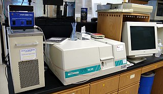
UV spectroscopy or UV–visible spectrophotometry refers to absorption spectroscopy or reflectance spectroscopy in part of the ultraviolet and the full, adjacent visible regions of the electromagnetic spectrum. Being relatively inexpensive and easily implemented, this methodology is widely used in diverse applied and fundamental applications. The only requirement is that the sample absorb in the UV-Vis region, i.e. be a chromophore. Absorption spectroscopy is complementary to fluorescence spectroscopy. Parameters of interest, besides the wavelength of measurement, are absorbance (A) or transmittance (%T) or reflectance (%R), and its change with time.

The enzyme-linked immunosorbent assay (ELISA) is a commonly used analytical biochemistry assay, first described by Eva Engvall and Peter Perlmann in 1971. The assay uses a solid-phase type of enzyme immunoassay (EIA) to detect the presence of a ligand in a liquid sample using antibodies directed against the protein to be measured. ELISA has been used as a diagnostic tool in medicine, plant pathology, and biotechnology, as well as a quality control check in various industries.

Scattering is a term used in physics to describe a wide range of physical processes where moving particles or radiation of some form, such as light or sound, are forced to deviate from a straight trajectory by localized non-uniformities in the medium through which they pass. In conventional use, this also includes deviation of reflected radiation from the angle predicted by the law of reflection. Reflections of radiation that undergo scattering are often called diffuse reflections and unscattered reflections are called specular (mirror-like) reflections. Originally, the term was confined to light scattering. As more "ray"-like phenomena were discovered, the idea of scattering was extended to them, so that William Herschel could refer to the scattering of "heat rays" in 1800. John Tyndall, a pioneer in light scattering research, noted the connection between light scattering and acoustic scattering in the 1870s. Near the end of the 19th century, the scattering of cathode rays and X-rays was observed and discussed. With the discovery of subatomic particles and the development of quantum theory in the 20th century, the sense of the term became broader as it was recognized that the same mathematical frameworks used in light scattering could be applied to many other phenomena.

A nephelometer or aerosol photometer is an instrument for measuring the concentration of suspended particulates in a liquid or gas colloid. A nephelometer measures suspended particulates by employing a light beam and a light detector set to one side of the source beam. Particle density is then a function of the light reflected into the detector from the particles. To some extent, how much light reflects for a given density of particles is dependent upon properties of the particles such as their shape, color, and reflectivity. Nephelometers are calibrated to a known particulate, then use environmental factors (k-factors) to compensate lighter or darker colored dusts accordingly. K-factor is determined by the user by running the nephelometer next to an air sampling pump and comparing results. There are a wide variety of research-grade nephelometers on the market as well as open source varieties.

An automated analyser is a medical laboratory instrument designed to measure different chemicals and other characteristics in a number of biological samples quickly, with minimal human assistance. These measured properties of blood and other fluids may be useful in the diagnosis of disease.

Absorption spectroscopy refers to spectroscopic techniques that measure the absorption of radiation, as a function of frequency or wavelength, due to its interaction with a sample. The sample absorbs energy, i.e., photons, from the radiating field. The intensity of the absorption varies as a function of frequency, and this variation is the absorption spectrum. Absorption spectroscopy is performed across the electromagnetic spectrum.
An assay is an investigative (analytic) procedure in laboratory medicine, mining, pharmacology, environmental biology and molecular biology for qualitatively assessing or quantitatively measuring the presence, amount, or functional activity of a target entity. The measured entity is often called the analyte, the measurand, or the target of the assay. The analyte can be a drug, biochemical substance, chemical element or compound, or cell in an organism or organic sample. An assay usually aims to measure an analyte's intensive property and express it in the relevant measurement unit.

Fluorescence spectroscopy is a type of electromagnetic spectroscopy that analyzes fluorescence from a sample. It involves using a beam of light, usually ultraviolet light, that excites the electrons in molecules of certain compounds and causes them to emit light; typically, but not necessarily, visible light. A complementary technique is absorption spectroscopy. In the special case of single molecule fluorescence spectroscopy, intensity fluctuations from the emitted light are measured from either single fluorophores, or pairs of fluorophores.
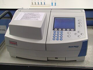
Plate readers, also known as microplate readers or microplate photometers, are instruments which are used to detect biological, chemical or physical events of samples in microtiter plates. They are widely used in research, drug discovery, bioassay validation, quality control and manufacturing processes in the pharmaceutical and biotechnological industry and academic organizations. Sample reactions can be assayed in 1-1536 well format microtiter plates. The most common microplate format used in academic research laboratories or clinical diagnostic laboratories is 96-well with a typical reaction volume between 100 and 200 µL per well. Higher density microplates are typically used for screening applications, when throughput and assay cost per sample become critical parameters, with a typical assay volume between 5 and 50 µL per well. Common detection modes for microplate assays are absorbance, fluorescence intensity, luminescence, time-resolved fluorescence, and fluorescence polarization.

The Tyndall effect is light scattering by particles in a colloid or in a very fine suspension. Also known as Tyndall scattering, it is similar to Rayleigh scattering, in that the intensity of the scattered light is inversely proportional to the fourth power of the wavelength, so blue light is scattered much more strongly than red light. An example in everyday life is the blue colour sometimes seen in the smoke emitted by motorcycles, in particular two-stroke machines where the burnt engine oil provides these particles.
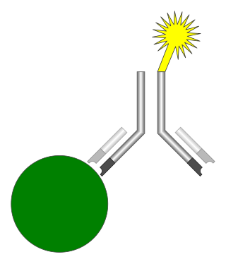
An immunoassay (IA) is a biochemical test that measures the presence or concentration of a macromolecule or a small molecule in a solution through the use of an antibody (usually) or an antigen (sometimes). The molecule detected by the immunoassay is often referred to as an "analyte" and is in many cases a protein, although it may be other kinds of molecules, of different sizes and types, as long as the proper antibodies that have the required properties for the assay are developed. Analytes in biological liquids such as serum or urine are frequently measured using immunoassays for medical and research purposes.
In physics, absorption of electromagnetic radiation is how matter takes up a photon's energy — and so transforms electromagnetic energy into internal energy of the absorber. A notable effect is attenuation, or the gradual reduction of the intensity of light waves as they propagate through a medium. Although the absorption of waves does not usually depend on their intensity, in certain conditions (optics) the medium's transparency changes by a factor that varies as a function of wave intensity, and saturable absorption occurs.

The Spectronic 20 is a brand of single-beam spectrophotometer, designed to operate in the visible spectrum across a wavelength range of 340 nm to 950 nm, with a spectral bandpass of 20 nm. It is designed for quantitative absorption measurement at single wavelengths. Because it measures the transmittance or absorption of visible light through a solution, it is sometimes referred to as a colorimeter. The name of the instrument is a trademark of the manufacturer.
Nephelometry is a technique used in immunology to determine the levels of several blood plasma proteins. For example the total levels of antibodies isotypes or classes: Immunoglobulin M, Immunoglobulin G, and Immunoglobulin A. It is important in quantification of free light chains in diseases such as multiple myeloma. Quantification is important for disease classification and for disease monitoring once a patient has been treated.
The photoacoustic Doppler effect is a type of Doppler effect that occurs when an intensity modulated light wave induces a photoacoustic wave on moving particles with a specific frequency. The observed frequency shift is a good indicator of the velocity of the illuminated moving particles. A potential biomedical application is measuring blood flow.

In physical and analytical chemistry, colorimetry or colourimetry is a technique used to determine the concentration of colored compounds in solution. A colorimeter is a device used to test the concentration of a solution by measuring its absorbance of a specific wavelength of light.
OD600 (Also written as O.D. 600, D600, o.d. 600, OD600) is an abbreviation indicating the optical density of a sample measured at a wavelength of 600 nm. It is a commonly used in Spectrophotometry for estimating the concentration of bacteria or other cells in a liquid as the 600nm wavelength does little to damage or hinder their growth. Optical density is largely based on light scattering and should not be confused with absorption.

Fluorescence polarization immunoassay (FPIA) is a class of in vitro biochemical test used for rapid detection of antibody or antigen in sample. FPIA is a competitive homogenous assay, that consists of a simple prepare and read method, without the requirement of separation or washing steps.
The collimated transmission method is a direct way of measuring the optical properties of materials. It is especially useful for sensing the optical properties of tissues to guide developments of both diagnostic and therapeutic techniques. These optical properties are described by the absorption coefficient μa, scattering coefficient μs, and anisotropy factor g.
References
- ↑ Mary C. Haven; Gregory A. Tetrault; Jerald R. Schenken (1994). Laboratory Instrumentation. John Wiley and Sons. ISBN 0471285722.
- ↑ D. M. Vasudevan; DM Vasudevan; S Sreekumari; Vaidyanathan Kannan (2010). Textbook of Biochemistry for Medical Students (6th ed.). Jaypee Medical Publishers. ISBN 978-9350250167.
| | This physical chemistry-related article is a stub. You can help Wikipedia by expanding it. |
| | This scattering–related article is a stub. You can help Wikipedia by expanding it. |