Tympanic membrane retraction

Last updated
Tympanic membrane retraction
Wiki TM retraction.jpg
Endoscopic image of retracted left tympanic membrane

Tympanic membrane retraction describes a condition in which a part of the eardrum lies deeper within the ear than its normal position.

Contents

The eardrum comprises two parts: the pars tensa, which is the main part of the eardrum, and the pars flaccida, which is a smaller part of the eardrum located above the pars tensa. Either or both of these parts may become retracted. The retracted segment of eardrum is often known as a retraction pocket. The terms atelectasis or sometimes adhesive otitis media can be used to describe retraction of a large area of the pars tensa.

Tympanic membrane retraction is fairly common and has been observed in one quarter of a population of British school children. [1] Retraction of both eardrums is less common than having a retraction in just one ear. It is more common in children with cleft palate. [2] Tympanic membrane retraction also occurs in adults. [3] [4]

Attempts have been made to categorise the extent of tympanic membrane retraction [3] [4] [5] though the validity of these classifications is limited. [2]

Presentation

The majority of tympanic membrane retractions do not cause any symptoms. Some cause hearing loss by restricting sound-induced vibrations of the eardrum. Permanent conductive hearing loss can be caused by erosion of the ossicles (hearing bones). Discharge from the ear often indicates that the retraction pocket has developed into a cholesteatoma.

Pathogenesis

Three factors must occur for the tympanic membrane to become retracted:

Negative middle ear pressure

When gas pressure within the middle ear is less than atmospheric pressure, the eardrum can become sucked into the middle ear space. This may be caused by disrupted gas exchange in the middle ear mucosa, inadequate opening of the Eustachian tube or a combination of these factors. People with a patulous Eustachian tube may also cause negative middle ear pressure by repeatedly sniffing to try to keep their Eustachian tube closed. [6]

Weakness of the tympanic membrane

The middle layer of the pars tensa is strengthened by fibers of collagen protein. This layer may be weaker in the postero-superior quadrant (top rear quarter) or after the eardrum heals after perforation or tympanostomy tubes (grommets) so predispose to retraction in these areas. The pars flaccida is prone to retraction as it does not contain the same stiffening layer of collagen.

Increase in surface area of the tympanic membrane

All over the body, new skin cells are continually produced to replace old skin cells which dry out and slough off. Growth of new cells on the surface of the eardrum is unusual in that the new cells migrate over the surface and move out along the ear canal. Even if migration along the ear canal is blocked, new cells continue to grow so the surface of the eardrum becomes larger. This process of proliferation and migration can result in enlargement of a retraction pocket so that the eardrum expands and grows deeper into the ear.

Natural history

The majority of tympanic membrane retractions remain stable for long periods of time, or may even resolve spontaneously so that the eardrum becomes normal again. [7] Not all retractions are able to resolve even if middle ear pressure normalizes, as the retracted segment may become adherent to other structures within the middle ear. Some retractions continue to progress and grow more deeply into the ear. This can result in erosion of bone and cholesteatoma formation. Asymptomatic deep pars tensa retractions tend to remain stable in adults. However, behaviour of symptomatic pars tensa retractions significantly differ between children and adults. There is a likelihood that such ears in children will recover spontaneously. There is no simple clinical means of identifying which ears will deteriorate to accumulate keratin debris in the pars tensa retraction, a phenomenon that is not influenced by age. In children, the pars tensa retraction may remain unchanged while the pars flaccida deteriorates to form cholesteatoma. [8]

Bone erosion

As the middle ear is only a narrow space, the eardrum only has to retract a short distance before it touches boney structures within the middle ear such as the ossicles. It may become adherent to these bones and in some cases, this contact leads to erosion of the bone. As well as ossicular erosion, the bone of the ear canal (e.g. the scutum) and even bone over the cochlea (the promontory) can become eroded.

Keratin entrapment

As skin cells die they form a barrier of dry protein called keratin. This layer of keratin is normally pushed out of the ear by migration of skin cells along the ear canal and is turned into wax. Clearance of keratin can be disrupted by tympanic membrane retraction so that keratin accumulates within the retraction pocket. When keratin becomes trapped deep inside the ear and cannot be cleaned out, it is known as cholesteatoma. Growth of bacteria in the trapped keratin causes smelly discharge from the ear and can spread to cause serious infection.

Treatment

Various strategies may be used to manage tympanic membrane retraction, with the aims of preventing or relieving hearing loss and cholesteatoma formation.

Observation

As retraction pockets may remain stable or resolve spontaneously, it may be appropriate to observe them for a period of time before considering any active treatment. [9]

Increase middle ear pressure

The Valsalva maneuver increases middle ear pressure and can push a retracted eardrum out of the middle ear if it is not adherent to middle ear structures. Hearing may improve as a result, however it can be a painful maneuver. The benefits are typically only temporary. Middle ear pressure can also be increased by Politzerization and with commercially available devices (e.g. Otovent and Ear Popper).

Ventilation tubes

A ventilation tube, also known as a tympanostomy tube or a grommet, may be placed through the eardrum to equalize middle ear pressure. Although this intervention may be effective, research has not yet shown whether it provides better results than simple observation. [10] Further weakness or perforation of the eardrum may occur.

Nasal treatments

These are intended to improve Eustachian tube function. Adenoidectomy can improve middle ear function [11] and nasal steroid sprays can reduce adenoid size [12] but it is not known whether these treatments alter tympanic membrane retraction.

Eustachian tube surgery

Enlargement of the Eustachian tube opening in the nose with laser or balloon dilation is being evaluated as a potential treatment for tympanic membrane retraction. [13] [14] There are two methods for this technique: Dennis Poe popularized the transnasal introduction of the balloon catheter to dilate the distal (nose end of the Eustachian tube). [14] Muaaz Tarabichi pioneered the dilatation of the proximal (ear side of the eustachian tube) through transtympanic (trans-ear) introduction of the balloon catheter. [15] [16]

Retraction pocket surgery

Excision of the retracted segment of an eardrum, with or without placement of a tympanostomy tube has been advocated. Healing of the area can result in a more normal eardrum. [17] Laser therapy has been used to shrink and stiffen retraction pockets. [18]

Tympanoplasty

Tympanoplasty is the surgical technique of removal of the retracted area from the middle ear and reconstruction of the tympanic membrane. Some surgeons use cartilage (taken from the outer ear) to stiffen the eardrum with the aim of preventing further retraction. [9] Surgical removal is required once a cholesteatoma has formed.

Related Research Articles

<span class="mw-page-title-main">Middle ear</span> Portion of the ear internal to the eardrum, and external to the oval window of the inner ear

The middle ear is the portion of the ear medial to the eardrum, and distal to the oval window of the cochlea.

<span class="mw-page-title-main">Cholesteatoma</span> Medical condition

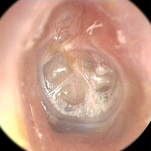
Cholesteatoma is a destructive and expanding growth consisting of keratinizing squamous epithelium in the middle ear and/or mastoid process. Cholesteatomas are not cancerous as the name may suggest, but can cause significant problems because of their erosive and expansile properties. This can result in the destruction of the bones of the middle ear (ossicles), as well as growth through the base of the skull into the brain. They often become infected and can result in chronically draining ears. Treatment almost always consists of surgical removal.

<span class="mw-page-title-main">Eardrum</span> Membrane separating the external ear from the middle ear

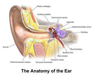
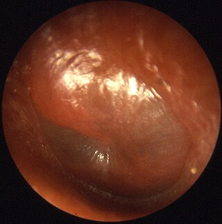
In the anatomy of humans and various other tetrapods, the eardrum, also called the tympanic membrane or myringa, is a thin, cone-shaped membrane that separates the external ear from the middle ear. Its function is to transmit sound from the air to the ossicles inside the middle ear, and then to the oval window in the fluid-filled cochlea. Hence, it ultimately converts and amplifies vibration in the air to vibration in cochlear fluid. The malleus bone bridges the gap between the eardrum and the other ossicles.

<span class="mw-page-title-main">Otitis media</span> Inflammation of the middle ear

Otitis media is a group of inflammatory diseases of the middle ear. One of the two main types is acute otitis media (AOM), an infection of rapid onset that usually presents with ear pain. In young children this may result in pulling at the ear, increased crying, and poor sleep. Decreased eating and a fever may also be present. The other main type is otitis media with effusion (OME), typically not associated with symptoms, although occasionally a feeling of fullness is described; it is defined as the presence of non-infectious fluid in the middle ear which may persist for weeks or months often after an episode of acute otitis media. Chronic suppurative otitis media (CSOM) is middle ear inflammation that results in a perforated tympanic membrane with discharge from the ear for more than six weeks. It may be a complication of acute otitis media. Pain is rarely present. All three types of otitis media may be associated with hearing loss. If children with hearing loss due to OME do not learn sign language, it may affect their ability to learn.

<span class="mw-page-title-main">Eustachian tube</span> Tube connecting middle ear to throat

The Eustachian tube, also called the auditory tube or pharyngotympanic tube, is a tube that links the nasopharynx to the middle ear, of which it is also a part. In adult humans, the Eustachian tube is approximately 35 mm (1.4 in) long and 3 mm (0.12 in) in diameter. It is named after the sixteenth-century Italian anatomist Bartolomeo Eustachi.

<span class="mw-page-title-main">Conductive hearing loss</span> Medical condition

Conductive hearing loss (CHL) occurs when there is a problem transferring sound waves anywhere along the pathway through the outer ear, tympanic membrane (eardrum), or middle ear (ossicles). If a conductive hearing loss occurs in conjunction with a sensorineural hearing loss, it is referred to as a mixed hearing loss. Depending upon the severity and nature of the conductive loss, this type of hearing impairment can often be treated with surgical intervention or pharmaceuticals to partially or, in some cases, fully restore hearing acuity to within normal range. However, cases of permanent or chronic conductive hearing loss may require other treatment modalities such as hearing aid devices to improve detection of sound and speech perception.

A myringotomy is a surgical procedure in which an incision is created in the eardrum to relieve pressure caused by excessive buildup of fluid, or to drain pus from the middle ear. A tympanostomy tube may be inserted through the eardrum to keep the middle ear aerated for a prolonged time and to prevent reaccumulation of fluid. Without the insertion of a tube, the incision usually heals spontaneously within two to three weeks. Depending on the type, the tube is either naturally extruded in 6 to 12 months or removed during a minor procedure.

<span class="mw-page-title-main">Tympanoplasty</span> Surgical operation on the ear

Tympanoplasty is the surgical operation performed to reconstruct hearing mechanism of middle ear.

<span class="mw-page-title-main">Tympanostomy tube</span> Medical device inserted into the eardrum

Tympanostomy tube, also known as a grommet or myringotomy tube, is a small tube inserted into the eardrum in order to keep the middle ear aerated for a prolonged period of time, and to prevent the accumulation of fluid in the middle ear. The operation to insert the tube involves a myringotomy and is performed under local or general anesthesia. The tube itself is made in a variety of designs. The most commonly used type is shaped like a grommet. When it is necessary to keep the middle ear ventilated for a very long period, a T-shaped tube may be used, as these "T-tubes" can stay in place for 2–4 years. Materials used to construct the tube are most often plastics such as silicone or Teflon. Stainless steel tubes exist, but are no longer in frequent use.

<span class="mw-page-title-main">Mastoiditis</span> Middle ear disease

Mastoiditis is the result of an infection that extends to the air cells of the skull behind the ear. Specifically, it is an inflammation of the mucosal lining of the mastoid antrum and mastoid air cell system inside the mastoid process. The mastoid process is the portion of the temporal bone of the skull that is behind the ear. The mastoid process contains open, air-containing spaces. Mastoiditis is usually caused by untreated acute otitis media and used to be a leading cause of child mortality. With the development of antibiotics, however, mastoiditis has become quite rare in developed countries where surgical treatment is now much less frequent and more conservative, unlike former times.

<span class="mw-page-title-main">Tympanometry</span>

Tympanometry is an acoustic evaluation of the condition of the middle ear eardrum and the conduction bones by creating variations of air pressure in the ear canal.

<span class="mw-page-title-main">Perforated eardrum</span> Injury leading to a hole in the eardrum

A perforated earhole is a prick in the eardrum. It can be caused by infection, trauma, overpressure, inappropriate ear clearing, and changes in middle ear pressure. An otoscope can be used to view the eardrum to diagnose a perforation. Perforations may heal naturally or require surgery.

<span class="mw-page-title-main">Prussak's space</span>

In human anatomy, Prussak's space is the small middle ear recess, bordered laterally by the flaccid part of Shrapnell's membrane, superiorly by the scutum and lateral malleal ligament, inferiorly by the lateral process of the malleus, and medially by the neck of the malleus. From the neck of the malleus, the anterior malleolar fold and the anterior ligament arise, demarcating Prussak's space anteriorly. Ventilation of Prussak's space is only possible posteriorly above the posterior malleus fold.

A mastoidectomy is a procedure performed to remove the mastoid air cells, air bubbles in the skull, near the inner ears. This can be done as part of treatment for mastoiditis, chronic suppurative otitis media or cholesteatoma. In addition, it is sometimes performed as part of other procedures or for access to the middle ear. There are classically 5 different types of mastoidectomy:

Myringoplasty is the closure of the perforation of pars tensa of the tympanic membrane. When myringoplasty is combined with removal of scar tissue, it is called tympanoplasty. The operation is performed with the patient supine and face turned to one side. The graft material most commonly used for the surgery is temporalis fascia. The tragal cartilage and tragal perichondrium are also used as the graft by some surgeons.

<span class="mw-page-title-main">Tympanosclerosis</span> Medical condition

Tympanosclerosis is a condition caused by hyalinization and subsequent calcification of subepithelial connective tissue of the tympanic membrane and middle ear, sometimes resulting in a detrimental effect to hearing.

Endoscopic ear surgery (EES) is a minimally invasive alternative to traditional ear surgery and is defined as the use of the rigid endoscope, as opposed to a surgical microscope, to visualize the middle and inner ear during otologic surgery. During endoscopic ear surgery the surgeon holds the endoscope in one hand while working in the ear with the other. To allow this kind of single-handed surgery, different surgical instruments have to be used. Endoscopic visualization has improved due to high-definition video imaging and wide-field endoscopy, and being less invasive, EES is gaining importance as an adjunct to microscopic ear surgery.

<span class="mw-page-title-main">Jacob Sadé</span> German-born Israeli otolaryngologist (1925–2020)

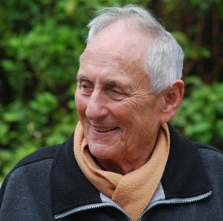
Jacob Sadé (3 June 1925 – 16 March 2020) was a German-born Israeli otolaryngologist, also an emeritus professor at the Sackler School of Medicine, Tel Aviv University.

Eustachian tube dysfunction (ETD) is a disorder where pressure abnormalities in the middle ear result in symptoms.

Muaaz Tarabichi is a Syrian otolaryngologist, lecturer, researcher, and author. He is recognized around the world as the father of endoscopic ear surgery. He is the co-founder of Tarabichi Stammberger Ear and Sinus Institute. He was elected as the chairman of the International Advisory Board of the American Academy of Otolaryngology–Head and Neck Surgery.

References

  1. Maw, AR; Hall AJ; Pothier DD; Gregory SP; Steer CD. (2011). "The prevalence of tympanic membrane and related middle ear pathology in children: a large longitudinal cohort study followed from birth to age ten". Otology & Neurotology. 32 (8): 1256–61. doi:10.1097/mao.0b013e31822f10cf. PMID   21897314. S2CID   13913918.
  2. 1 2 James AL, Papsin BC, Trimble K, Ramsden J, Sanjeevan N, Bailie N, Chadha NK (May 2012). "Tympanic membrane retraction: An endoscopic evaluation of staging systems". Laryngoscope. 122 (5): 1115–20. doi:10.1002/lary.23203. PMID   22374833. S2CID   6122719.
  3. 1 2 Sadé, J; Berco E (1976). "Atelectasis and secretory otitis media". Ann Otol Rhinol Laryngol. 85 (2 Suppl 25 Pt 2): 66–72. doi:10.1177/00034894760850S214. PMID   1267370. S2CID   42460109.
  4. 1 2 Tos, M; Poulsen G (1980). "Attic retractions following secretory otitis". Acta Otolaryngol. 89 (5–6): 479–86. doi:10.3109/00016488009127165. PMID   7192477.
  5. Borgstein, J; Gerritsma TV; Wieringa MH; Bruce IA (2007). "The Erasmus atelectasis classification: proposal of a new classification for atelectasis of the middle ear in children". Laryngoscope. 117 (7): 1255–9. doi:10.1097/mlg.0b013e31805d0160. PMID   17603325. S2CID   21170363.
  6. Ikeda, R; Oshima T; Oshima H; Miyazaki M; Kikuchi T; Kawase T; Kobayashi T (2011). "Management of patulous Eustachian tube with habitual sniffing". Otology & Neurotology. 32 (5): 790–3. doi:10.1097/mao.0b013e3182184e23. PMID   21659936. S2CID   25034104.
  7. MRC Multi-Centre Otitis Media Study Group (May 2001). "Pars tensa and pars flaccida retractions in persistent otitis media with effusion". Otol. Neurotol. 22 (3): 291–8. doi:10.1097/00129492-200105000-00004. PMID   11347629. S2CID   11338999.
  8. John Cutajar, Maryam Nowghani, Bharti Tulsidas-Mahtani, and John Hamilton (April 2018). "The Natural History of Asymptomatic Deep Pars Tensa Retraction". Int. Adv. Otol. 14 (1): 10–14. doi: 10.5152/iao.2018.5234 . PMC   6354506 . PMID   29764774.{{cite journal}}: CS1 maint: multiple names: authors list (link)
  9. 1 2 Bluestone, Charles (2005). Eustachian Tube. Hamilton, ON: B C Decker Inc. pp. 189–192. ISBN   978-1-55009-066-6.
  10. Nankivell, PC; Pothier DD (7 Jul 2010). "Surgery for tympanic membrane retraction pockets". Cochrane Database Syst Rev. 7 (7): CD007943. doi:10.1002/14651858.CD007943.pub2. PMID   20614467.
  11. van den Aardweg, MT; Schilder AG; Herkert E; Boonacker CW; Rovers MM (20 January 2010). "Adenoidectomy for otitis media in children". Cochrane Database Syst Rev. CD007810 (1): CD007810. doi:10.1002/14651858.CD007810.pub2. PMID   20091650.
  12. Zhang, L; Mendoza-Sassi RA; César JA; Chadha NK (16 July 2008). "Intranasal corticosteroids for nasal airway obstruction in children with moderate to severe adenoidal hypertrophy". Cochrane Database Syst Rev. 3 (3): CD006286. doi:10.1002/14651858.CD006286.pub2. PMC   8923350 . PMID   18646145.
  13. Poe, DS; Silvola J; Pyykkö I (2011). "Balloon dilation of the cartilaginous eustachian tube". Otolaryngol Head Neck Surg. 144 (4): 563–9. doi:10.1177/0194599811399866. PMID   21493236. S2CID   10717771.
  14. 1 2 Poe, Dennis; Anand, Vijay; Dean, Marc; Roberts, William H.; Stolovitzky, Jose Pablo; Hoffmann, Karen; Nachlas, Nathan E.; Light, Joshua P.; Widick, Mark H.; Sugrue, John P.; Elliott, C. Layton (May 2018). "Balloon dilation of the eustachian tube for dilatory dysfunction: A randomized controlled trial: Balloon Dilation of the Eustachian Tube". The Laryngoscope. 128 (5): 1200–1206. doi: 10.1002/lary.26827 . PMID   28940574. S2CID   4968887.
  15. Tarabichi, Muaaz; Najmi, Murtaza (2015-07-03). "Transtympanic dilatation of the eustachian tube during chronic ear surgery". Acta Oto-Laryngologica. 135 (7): 640–644. doi:10.3109/00016489.2015.1009640. ISSN   0001-6489. PMID   25762371. S2CID   39239009.
  16. Kapadia, Mustafa; Tarabichi, Muaaz (October 2018). "Feasibility and Safety of Transtympanic Balloon Dilatation of Eustachian Tube". Otology & Neurotology. 39 (9): e825–e830. doi:10.1097/MAO.0000000000001950. ISSN   1531-7129. PMID   30124616. S2CID   52041093.
  17. Blaney, SP; Tierney P; Bowdler DA (1999). "The surgical management of the pars tensa retraction pocket in the child--results following simple excision and ventilation tube insertion". Int J Pediatr Otorhinolaryngol. 50 (2): 133–7. doi:10.1016/s0165-5876(99)00227-x. PMID   10576614.
  18. Brawner, JT; Saunders JE; Berryhill WE. (2008). "Laser myringoplasty for tympanic membrane atelectasis". Otolaryngol Head Neck Surg. 139 (1): 47–50. doi:10.1016/j.otohns.2008.01.018. PMID   18585560. S2CID   2457206.