Tympanosclerosis

Last updated
Tympanoesclerosis
Other namesMyringosclerosis, intratympanic tympanosclerosis
Tympanosclerosis.jpg
Specialty ENT surgery

Tympanosclerosis is a condition caused by hyalinization and subsequent calcification of subepithelial connective tissue of the tympanic membrane and middle ear, sometimes resulting in a detrimental effect to hearing. [1] [2]

Contents

Signs and symptoms

Myringosclerosis rarely causes any symptoms. [3] Tympanosclerosis, on the other hand, can cause significant hearing loss [3] or chalky, white patches on the middle ear or tympanic membrane. [1]

Causes

The aetiology for tympanosclerosis is not extensively understood. There are several probable factors which could result in the condition appearing, including:

Pathophysiology

Increased fibroblast activity results in deposition of collagen. Calcium phosphate plaques then form in the lamina propria of the tympanic membrane. [1] [2]

Diagnosis

If lesions are typical, non-extensive and with no detriment to hearing, investigation into the condition is rarely required. Audiometry is used to determine the extent of hearing loss, if any. [3] Tympanometry produces tympanograms which can be different when tympanosclerosis is present. [11] Computerised tomography (CT) can be used to determine if disease is present in the middle ear. [12] Whilst hearing loss is a common symptom in many diseases of the ear, for example in otosclerosis (abnormal bone growth in the ear), [3] the white, chalky patches on the tympanic membrane are fairly characteristic of tympanosclerosis. Cholesteatoma is similar in appearance but the whiteness is behind the tympanic membrane, rather than inside.[ citation needed ]

Classification

Myringosclerosis refers to a calcification only within the tympanic membrane and is usually less extensive than intratympanic tympanosclerosis, which refers to any other location within the middle ear such as the ossicular chain, middle ear mucosa or, less frequently, the mastoid cavity. [3]

Treatment

Hearing aids are a common treatment for hearing loss disorders. A more specific treatment is surgical, involving excision of the sclerotic areas and then further repair of the ossicular chain. There are several techniques, sometimes involving two surgeries; [2] [13] success rates are, however, variable. [14] Damage to the inner ear as a result of surgical procedures is a possible and serious concern, as it can result in forms of sensorineural deafness. [15]

Prognosis

In most cases, tympanosclerosis does not cause any recognisable hearing loss up to ten years after the initial disease onset. [5] [16] Sclerotic changes seem to stabilise, but not resolve or dissolve, after 3 years. [9] [17]

Epidemiology

Myringosclerosis seems to be more common than tympanosclerosis. [2] Most research has not been conducted upon the general, healthy population, but rather those with otitis media or patients who have had tympanostomy tubes in prior procedures. Of the children studied who had 'glue ear', and who were treated with tympanostomy tubing, 23-40% of cases had tympanosclerosis. [4] [9] [18] One study suggested that people with atherosclerosis were more likely to have tympanosclerosis than otherwise healthy individuals. [10]

Related Research Articles

<span class="mw-page-title-main">Cholesteatoma</span> Medical condition

Cholesteatoma is a destructive and expanding growth consisting of keratinizing squamous epithelium in the middle ear and/or mastoid process. Cholesteatomas are not cancerous as the name may suggest, but can cause significant problems because of their erosive and expansile properties. This can result in the destruction of the bones of the middle ear (ossicles), as well as growth through the base of the skull into the brain. They often become infected and can result in chronically draining ears. Treatment almost always consists of surgical removal.

<span class="mw-page-title-main">Eardrum</span> Membrane separating the external ear from the middle ear

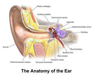
In the anatomy of humans and various other tetrapods, the eardrum, also called the tympanic membrane or myringa, is a thin, cone-shaped membrane that separates the external ear from the middle ear. Its function is to transmit sound from the air to the ossicles inside the middle ear, and then to the oval window in the fluid-filled cochlea. Hence, it ultimately converts and amplifies vibration in the air to vibration in cochlear fluid. The malleus bone bridges the gap between the eardrum and the other ossicles.

<span class="mw-page-title-main">Otitis media</span> Inflammation of the middle ear

Otitis media is a group of inflammatory diseases of the middle ear. One of the two main types is acute otitis media (AOM), an infection of rapid onset that usually presents with ear pain. In young children this may result in pulling at the ear, increased crying, and poor sleep. Decreased eating and a fever may also be present. The other main type is otitis media with effusion (OME), typically not associated with symptoms, although occasionally a feeling of fullness is described; it is defined as the presence of non-infectious fluid in the middle ear which may persist for weeks or months often after an episode of acute otitis media. Chronic suppurative otitis media (CSOM) is middle ear inflammation that results in a perforated tympanic membrane with discharge from the ear for more than six weeks. It may be a complication of acute otitis media. Pain is rarely present. All three types of otitis media may be associated with hearing loss. If children with hearing loss due to OME do not learn sign language, it may affect their ability to learn.

<span class="mw-page-title-main">Eustachian tube</span> Tube connecting middle ear to throat

The Eustachian tube, also called the auditory tube or pharyngotympanic tube, is a tube that links the nasopharynx to the middle ear, of which it is also a part. In adult humans, the Eustachian tube is approximately 35 mm (1.4 in) long and 3 mm (0.12 in) in diameter. It is named after the sixteenth-century Italian anatomist Bartolomeo Eustachi.

<span class="mw-page-title-main">Otosclerosis</span> Condition characterized by an abnormal bone growth in the middle ear

Otosclerosis is a condition of the middle ear where portions of the dense enchondral layer of the bony labyrinth remodel into one or more lesions of irregularly-laid spongy bone. As the lesions reach the stapes the bone is resorbed, then hardened (sclerotized), which limits its movement and results in hearing loss, tinnitus, vertigo or a combination of symptoms. The term otosclerosis is something of a misnomer: much of the clinical course is characterized by lucent rather than sclerotic bony changes, so the disease is also known as otospongiosis.

Stapedectomy is a surgical procedure in which the stapes bone is removed from the middle ear and replaced with a prosthesis.

<span class="mw-page-title-main">Conductive hearing loss</span> Medical condition

Conductive hearing loss (CHL) occurs when there is a problem transferring sound waves anywhere along the pathway through the outer ear, tympanic membrane (eardrum), or middle ear (ossicles). If a conductive hearing loss occurs in conjunction with a sensorineural hearing loss, it is referred to as a mixed hearing loss. Depending upon the severity and nature of the conductive loss, this type of hearing impairment can often be treated with surgical intervention or pharmaceuticals to partially or, in some cases, fully restore hearing acuity to within normal range. However, cases of permanent or chronic conductive hearing loss may require other treatment modalities such as hearing aid devices to improve detection of sound and speech perception.

A myringotomy is a surgical procedure in which an incision is created in the eardrum to relieve pressure caused by excessive buildup of fluid, or to drain pus from the middle ear. A tympanostomy tube may be inserted through the eardrum to keep the middle ear aerated for a prolonged time and to prevent reaccumulation of fluid. Without the insertion of a tube, the incision usually heals spontaneously within two to three weeks. Depending on the type, the tube is either naturally extruded in 6 to 12 months or removed during a minor procedure.

<span class="mw-page-title-main">Tympanoplasty</span> Surgical operation on the ear

Tympanoplasty is the surgical operation performed to reconstruct hearing mechanism of middle ear.

<span class="mw-page-title-main">Otology</span> Branch of medicine for the ear

Otology is a branch of medicine which studies normal and pathological anatomy and physiology of the ear as well as their diseases, diagnosis and treatment. Otologic surgery generally refers to surgery of the middle ear and mastoid related to chronic otitis media, such as tympanoplasty, or ear drum surgery, ossiculoplasty, or surgery of the hearing bones, and mastoidectomy. Otology also includes surgical treatment of conductive hearing loss, such as stapedectomy surgery for otosclerosis.

<span class="mw-page-title-main">Tympanostomy tube</span> Medical device inserted into the eardrum

Tympanostomy tube, also known as a grommet or myringotomy tube, is a small tube inserted into the eardrum in order to keep the middle ear aerated for a prolonged period of time, and to prevent the accumulation of fluid in the middle ear. The operation to insert the tube involves a myringotomy and is performed under local or general anesthesia. The tube itself is made in a variety of designs. The most commonly used type is shaped like a grommet. When it is necessary to keep the middle ear ventilated for a very long period, a "T"-shaped tube may be used, as these "T-tubes" can stay in place for 2–4 years. Materials used to construct the tube are most often plastics such as silicone or Teflon. Stainless steel tubes exist, but are no longer in frequent use.

Burow's solution is an aqueous solution of aluminium triacetate. It is available in the U.S. as an over-the-counter drug for topical administration, with brand names including Domeboro, Domeboro Otic, Star-Otic, and Borofair. The preparation has astringent and antibacterial properties and may be used to treat a number of skin conditions, including insect bites and stings, rashes caused by poison ivy and poison sumac, swelling, allergies, and bruises. However, its main use is for treatment of otitis, including otomycosis.

<span class="mw-page-title-main">Perforated eardrum</span> Injury leading to a hole in the eardrum

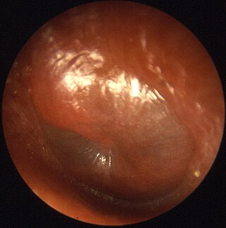
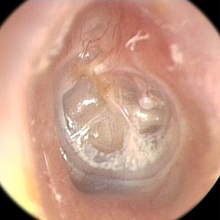
A perforated eardrum is a hole in the eardrum. It can be caused by infection, trauma, overpressure, inappropriate ear clearing, and changes in middle ear pressure. An otoscope can be used to view the eardrum to diagnose a perforation. Perforations may heal naturally or require surgery.

Hearing loss with craniofacial syndromes is a common occurrence. Many of these multianomaly disorders involve structural malformations of the outer or middle ear, making a significant hearing loss highly likely.

A mastoidectomy is a procedure performed to remove the mastoid air cells, air bubbles in the skull, near the inner ears. This can be done as part of treatment for mastoiditis, chronic suppurative otitis media or cholesteatoma. In addition, it is sometimes performed as part of other procedures or for access to the middle ear. There are classically 5 different types of mastoidectomy:

<span class="mw-page-title-main">Tympanic membrane retraction</span> Medical condition

Tympanic membrane retraction describes a condition in which a part of the eardrum lies deeper within the ear than its normal position.

<span class="mw-page-title-main">Otic polyp</span> Medical condition

An otic polyp is a benign proliferation of chronic inflammatory cells associated with granulation tissue, in response to a longstanding inflammatory process of the middle ear.

Nicholas John Frootko is a retired South African / British Otolaryngologist / Head and Neck Surgeon with a special interest and expertise in Ear Surgery.

Eustachian tube dysfunction (ETD) is a disorder where pressure abnormalities in the middle ear result in symptoms.

A middle ear implant is a hearing device that is surgically implanted into the middle ear. They help people with conductive, sensorineural or mixed hearing loss to hear. 

References

  1. 1 2 3 Forséni M, Bagger-Sjöbäck D, Hultcrantz M (May 2001). "A study of inflammatory mediators in the human tympanosclerotic middle ear". Archives of Otolaryngology–Head & Neck Surgery. 127 (5): 559–64. doi: 10.1001/archotol.127.5.559 . PMID   11346433. Archived from the original on 2011-08-10. Retrieved 2010-01-26.
  2. 1 2 3 4 5 6 Asiri S, Hasham A, al Anazy F, Zakzouk S, Banjar A (December 1999). "Tympanosclerosis: review of literature and incidence among patients with middle-ear infection". The Journal of Laryngology and Otology. 113 (12): 1076–80. doi:10.1017/s0022215100157937. PMID   10767919.
  3. 1 2 3 4 5 Isaacson JE, Vora NM (September 2003). "Differential diagnosis and treatment of hearing loss". American Family Physician. 68 (6): 1125–32. PMID   14524400.
  4. 1 2 Kay DJ, Nelson M, Rosenfeld RM (April 2001). "Meta-analysis of tympanostomy tube sequelae". Otolaryngology–Head and Neck Surgery . 124 (4): 374–80. doi:10.1067/mhn.2001.113941. PMID   11283489.
  5. 1 2 Johnston LC, Feldman HM, Paradise JL, et al. (July 2004). "Tympanic membrane abnormalities and hearing levels at the ages of 5 and 6 years in relation to persistent otitis media and tympanostomy tube insertion in the first 3 years of life: a prospective study incorporating a randomized clinical trial". Pediatrics. 114 (1): e58–67. doi: 10.1542/peds.114.1.e58 . PMID   15231974 . Retrieved 2010-01-26.
  6. Browning, George G.; Rovers, Maroeska M.; Williamson, Ian; Lous, Jørgen; Burton, Martin J. (2010-10-06). "Grommets (ventilation tubes) for hearing loss associated with otitis media with effusion in children". The Cochrane Database of Systematic Reviews (10): CD001801. doi:10.1002/14651858.CD001801.pub3. ISSN   1469-493X. PMID   20927726.
  7. McRae D, Gatland DJ, Youngs R, Cook J (August 1989). "Aspiration of middle ear effusions prior to grommet insertion an etiological factor in tympanosclerosis". The Journal of Otolaryngology. 18 (5): 229–31. PMID   2769837.
  8. Hampal S, Flood LM, Kumar BU (March 1991). "The mini-grommet and tympanosclerosis". The Journal of Laryngology and Otology. 105 (3): 161–4. doi:10.1017/s0022215100115269. PMID   2019798.
  9. 1 2 3 Maw AR (August 1991). "Development of tympanosclerosis in children with otitis media with effusion and ventilation tubes". The Journal of Laryngology and Otology. 105 (8): 614–7. doi:10.1017/s0022215100116822. PMID   1919311.
  10. 1 2 Ferri M, Faggioli GL, Ferri GG, Pirodda A (June 2004). "Is carotid stenosis correlated with tympanosclerosis". International Angiology. 23 (2): 144–6. PMID   15507892 . Retrieved 2010-01-26.
  11. Onusko E (November 2004). "Tympanometry". American Family Physician. 70 (9): 1713–20. PMID   15554489.
  12. Swartz JD, Goodman RS, Russell KB, Marlowe FI, Wolfson RJ (August 1983). "High-resolution computed tomography of the middle ear and mastoid. Part II: Tubotympanic disease". Radiology. 148 (2): 455–9. doi:10.1148/radiology.148.2.6867342. PMID   6867342.
  13. Bayazit YA, Ozer E, Kara C, Gökpinar S, Kanlikama M, Mumbuç S (May 2004). "An analysis of the single-stage tympanoplasty with over-underlay grafting in tympanosclerosis". Otology & Neurotology. 25 (3): 211–4. doi:10.1097/00129492-200405000-00001. PMID   15129093.
  14. Vincent R, Oates J, Sperling NM (November 2002). "Stapedotomy for tympanosclerotic stapes fixation: is it safe and efficient? A review of 68 cases". Otology & Neurotology. 23 (6): 866–72. doi:10.1097/00129492-200211000-00010. PMID   12438848.
    - Albu S, Babighian G, Trabalzini F (September 2000). "Surgical treatment of tympanosclerosis". The American Journal of Otology. 21 (5): 631–5. PMID   10993449.
    - Teufert KB, De La Cruz A (March 2002). "Tympanosclerosis: long-term hearing results after ossicular reconstruction". Otolaryngology–Head and Neck Surgery . 126 (3): 264–72. doi:10.1067/mhn.2002.122701. PMID   11956534.
  15. Bellucci RJ (August 1985). "Cochlear hearing loss in tympanoplasty". Otolaryngology–Head and Neck Surgery . 93 (4): 482–5. doi:10.1177/019459988509300403. PMID   3931022.
  16. Riley DN, Herberger S, McBride G, Law K (March 1997). "Myringotomy and ventilation tube insertion: a ten-year follow-up". The Journal of Laryngology and Otology. 111 (3): 257–61. doi:10.1017/s0022215100137016. PMID   9156062.
  17. De Beer BA, Schilder AG, Zielhuis GA, Graamans K (September 2005). "Natural course of tympanic membrane pathology related to otitis media and ventilation tubes between ages 8 and 18 years". Otology & Neurotology. 26 (5): 1016–21. doi:10.1097/01.mao.0000185058.89586.ed. PMID   16151352.
  18. Pereira MB, Pereira DR, Costa SS (2005). "Tympanostomy tube sequelae in children with otitis media with effusion: a three-year follow-up study". Brazilian Journal of Otorhinolaryngology. 71 (4): 415–20. doi: 10.1590/S0034-72992005000400003 . hdl: 10183/48960 . PMID   16446953.
    - Schilder AG, Zielhuis GA, Haggard MP, van den Broek P (May 1995). "Long-term effects of otitis media with effusion: otomicroscopic findings". The American Journal of Otology. 16 (3): 365–72. PMID   8588632.