Related Research Articles

The gastrointestinal tract is the tract or passageway of the digestive system that leads from the mouth to the anus. The GI tract contains all the major organs of the digestive system, in humans and other animals, including the esophagus, stomach, and intestines. Food taken in through the mouth is digested to extract nutrients and absorb energy, and the waste expelled at the anus as faeces. Gastrointestinal is an adjective meaning of or pertaining to the stomach and intestines.

The ileum is the final section of the small intestine in most higher vertebrates, including mammals, reptiles, and birds. In fish, the divisions of the small intestine are not as clear and the terms posterior intestine or distal intestine may be used instead of ileum. Its main function is to absorb vitamin B12, bile salts, and whatever products of digestion that were not absorbed by the jejunum.
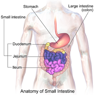
The small intestine or small bowel is an organ in the gastrointestinal tract where most of the absorption of nutrients from food takes place. It lies between the stomach and large intestine, and receives bile and pancreatic juice through the pancreatic duct to aid in digestion. The small intestine is about 5.5 metres long and folds many times to fit in the abdomen. Although it is longer than the large intestine, it is called the small intestine because it is narrower in diameter.

In biology, tissue is a historically derived biological organizational level between cells and a complete organ. A tissue is therefore often thought of as an assembly of similar cells and their extracellular matrix from the same embryonic origin that together carry out a specific function. Organs are then formed by the functional grouping together of multiple tissues.
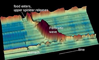
Peristalsis is a type of intestinal motility, characterized by radially symmetrical contraction and relaxation of muscles that propagate in a wave down a tube, in an anterograde direction. Peristalsis is progression of coordinated contraction of involuntary circular muscles, which is preceded by a simultaneous contraction of the longitudinal muscle and relaxation of the circular muscle in the lining of the gut.

Microvilli are microscopic cellular membrane protrusions that increase the surface area for diffusion and minimize any increase in volume, and are involved in a wide variety of functions, including absorption, secretion, cellular adhesion, and mechanotransduction.
Digestion is the breakdown of large insoluble food compounds into small water-soluble components so that they can be absorbed into the blood plasma. In certain organisms, these smaller substances are absorbed through the small intestine into the blood stream. Digestion is a form of catabolism that is often divided into two processes based on how food is broken down: mechanical and chemical digestion. The term mechanical digestion refers to the physical breakdown of large pieces of food into smaller pieces which can subsequently be accessed by digestive enzymes. Mechanical digestion takes place in the mouth through mastication and in the small intestine through segmentation contractions. In chemical digestion, enzymes break down food into the small compounds that the body can use.

Fish anatomy is the study of the form or morphology of fish. It can be contrasted with fish physiology, which is the study of how the component parts of fish function together in the living fish. In practice, fish anatomy and fish physiology complement each other, the former dealing with the structure of a fish, its organs or component parts and how they are put together, such as might be observed on the dissecting table or under the microscope, and the latter dealing with how those components function together in living fish.

In anatomy, the meninges are the three membranes that envelop the brain and spinal cord. In mammals, the meninges are the dura mater, the arachnoid mater, and the pia mater. Cerebrospinal fluid is located in the subarachnoid space between the arachnoid mater and the pia mater. The primary function of the meninges is to protect the central nervous system.

Intestinal villi are small, finger-like projections that extend into the lumen of the small intestine. Each villus is approximately 0.5–1.6 mm in length, and has many microvilli projecting from the enterocytes of its epithelium which collectively form the striated or brush border. Each of these microvilli are about 1 µm in length, around 1000 times shorter than a single villus. The intestinal villi are much smaller than any of the circular folds in the intestine.

Enterocytes, or intestinal absorptive cells, are simple columnar epithelial cells which line the inner surface of the small and large intestines. A glycocalyx surface coat contains digestive enzymes. Microvilli on the apical surface increase its surface area. This facilitates transport of numerous small molecules into the enterocyte from the intestinal lumen. These include broken down proteins, fats, and sugars, as well as water, electrolytes, vitamins, and bile salts. Enterocytes also have an endocrine role, secreting hormones such as leptin.
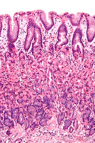
Simple columnar epithelium is a single layer of columnar epithelial cells which are tall and slender with oval-shaped nuclei located in the basal region, attached to the basement membrane. In humans, simple columnar epithelium lines most organs of the digestive tract including the stomach, and intestines. Simple columnar epithelium also lines the uterus.
Cyclostomi, often referred to as Cyclostomata, is a group of vertebrates that comprises the living jawless fishes: the lampreys and hagfishes. Both groups have jawless mouths with horny epidermal structures that function as teeth called ceratodontes, and branchial arches that are internally positioned instead of external as in the related jawed fishes. The name Cyclostomi means "round mouths". It was named by Joan Crockford-Beattie.
Absorption is the journey of a drug travelling from the site of administration to the site of action.

The intestinal epithelium is the single cell layer that form the luminal surface (lining) of both the small and large intestine (colon) of the gastrointestinal tract. Composed of simple columnar epithelial cells, it serves two main functions: absorbing useful substances into the body and restricting the entry of harmful substances. As part of its protective role, the intestinal epithelium forms an important component of the intestinal mucosal barrier. Certain diseases and conditions are caused by functional defects in the intestinal epithelium. On the other hand, various diseases and conditions can lead to its dysfunction which, in turn, can lead to further complications.

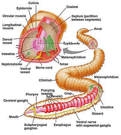
An earthworm is a soil-dwelling terrestrial invertebrate that belongs to the phylum Annelida. The term is the common name for the largest members of the class Oligochaeta. In classical systems, they were in the order of Opisthopora since the male pores opened posterior to the female pores, although the internal male segments are anterior to the female. Theoretical cladistic studies have placed them in the suborder Lumbricina of the order Haplotaxida, but this may change. Other slang names for earthworms include "dew-worm", "rainworm", "nightcrawler", and "angleworm". Larger terrestrial earthworms are also called megadriles as opposed to the microdriles in the semiaquatic families Tubificidae, Lumbricidae and Enchytraeidae. The megadriles are characterized by a distinct clitellum and a vascular system with true capillaries.
Microchaetus rappi, the African giant earthworm, is a large earthworm in the Microchaetidae family, the largest of the segmented worms. It averages about 1.4 meters in length, but can reach a length of as much as 6.7 meters and can weigh over 1.5 kilograms.

The gastrointestinal wall of the gastrointestinal tract is made up of four layers of specialised tissue. From the inner cavity of the gut outwards, these are:
- Mucosa
- Submucosa
- Muscular layer
- Serosa or adventitia

The human digestive system consists of the gastrointestinal tract plus the accessory organs of digestion. Digestion involves the breakdown of food into smaller and smaller components, until they can be absorbed and assimilated into the body. The process of digestion has three stages: the cephalic phase, the gastric phase, and the intestinal phase.

The pulmonary pleurae are the two opposing layers of serous membrane overlying the lungs, mediastinum and the inside surfaces of the surrounding chest walls.
References
- ↑ Fujii, Tamotsu (July 1982). "Electron microscopy of the leucocytes of the typhlosole in ammocoetes, with special attention to the antibody- producing cells". Journal of Morphology. 173 (1): 87–100. doi:10.1002/jmor.1051730108. ISSN 0362-2525. PMID 7050396. S2CID 37992278.
- ↑ "Typhlosole - an overview | ScienceDirect Topics". www.sciencedirect.com. Retrieved 2023-03-16.