Related Research Articles

X-ray is a form of high-energy electromagnetic radiation. In many languages, it is referred to as Röntgen radiation, after the German scientist Wilhelm Conrad Röntgen, who discovered it in 1895 and named it X-radiation to signify an unknown type of radiation.
In physics, attenuation is the gradual loss of flux intensity through a medium. For instance, dark glasses attenuate sunlight, lead attenuates X-rays, and water and air attenuate both light and sound at variable attenuation rates.

X-ray fluorescence (XRF) is the emission of characteristic "secondary" X-rays from a material that has been excited by being bombarded with high-energy X-rays or gamma rays. The phenomenon is widely used for elemental analysis and chemical analysis, particularly in the investigation of metals, glass, ceramics and building materials, and for research in geochemistry, forensic science, archaeology and art objects such as paintings.

Radiography is an imaging technique using X-rays, gamma rays, or similar ionizing radiation and non-ionizing radiation to view the internal form of an object. Applications of radiography include medical and industrial radiography. Similar techniques are used in airport security,. To create an image in conventional radiography, a beam of X-rays is produced by an X-ray generator and it is projected towards the object. A certain amount of the X-rays or other radiation are absorbed by the object, dependent on the object's density and structural composition. The X-rays that pass through the object are captured behind the object by a detector. The generation of flat two-dimensional images by this technique is called projectional radiography. In computed tomography, an X-ray source and its associated detectors rotate around the subject, which itself moves through the conical X-ray beam produced. Any given point within the subject is crossed from many directions by many different beams at different times. Information regarding the attenuation of these beams is collated and subjected to computation to generate two-dimensional images on three planes which can be further processed to produce a three-dimensional image.

External beam radiation therapy (EBRT) is a form of radiotherapy that utilizes a high-energy collimated beam of ionizing radiation, from a source outside the body, to target and kill cancer cells. A radiotherapy beam is composed of particles which travel in a consistent direction; each radiotherapy beam consists of one type of particle intended for use in treatment, though most beams contain some contamination by other particle types.

A monochromator is an optical device that transmits a mechanically selectable narrow band of wavelengths of light or other radiation chosen from a wider range of wavelengths available at the input. The name is from the Greek roots mono-, "single", and chroma, "colour", and the Latin suffix -ator, denoting an agent.
Wavelength-dispersive X-ray spectroscopy is a non-destructive analysis technique used to obtain elemental information about a range of materials by measuring characteristic x-rays within a small wavelength range. The technique generates a spectrum in which the peaks correspond to specific x-ray lines and elements can be easily identified. WDS is primarily used in chemical analysis, wavelength dispersive X-ray fluorescence (WDXRF) spectrometry, electron microprobes, scanning electron microscopes, and high precision experiments for testing atomic and plasma physics.
A collimator is a device which narrows a beam of particles or waves. To narrow can mean either to cause the directions of motion to become more aligned in a specific direction, or to cause the spatial cross section of the beam to become smaller.
X-ray optics is the branch of optics that manipulates X-rays instead of visible light. It deals with focusing and other ways of manipulating the X-ray beams for research techniques such as X-ray diffraction, X-ray crystallography, X-ray fluorescence, small-angle X-ray scattering, X-ray microscopy, X-ray phase-contrast imaging, and X-ray astronomy.

The Extreme Ultraviolet Explorer was a NASA space telescope for ultraviolet astronomy. EUVE was a part of NASA's Explorer spacecraft series. Launched on 7 June 1992. With instruments for ultraviolet (UV) radiation between wavelengths of 7 and 76 nm, the EUVE was the first satellite mission especially for the short-wave ultraviolet range. The satellite compiled an all-sky survey of 801 astronomical targets before being decommissioned on 31 January 2001.

X-ray diffraction is a generic term for phenomena associated with changes in the direction of X-ray beams due to interactions with the electrons around atoms. It occurs due to elastic scattering, when there is no change in the energy of the waves. The resulting map of the directions of the X-rays far from the sample is called a diffraction pattern. It is different from X-ray crystallography which exploits X-ray diffraction to determine the arrangement of atoms in materials, and also has other components such as ways to map from experimental diffraction measurements to the positions of atoms.

Projectional radiography, also known as conventional radiography, is a form of radiography and medical imaging that produces two-dimensional images by X-ray radiation. The image acquisition is generally performed by radiographers, and the images are often examined by radiologists. Both the procedure and any resultant images are often simply called 'X-ray'. Plain radiography or roentgenography generally refers to projectional radiography. Plain radiography can also refer to radiography without a radiocontrast agent or radiography that generates single static images, as contrasted to fluoroscopy, which are technically also projectional.

The mass attenuation coefficient, or mass narrow beam attenuation coefficient of a material is the attenuation coefficient normalized by the density of the material; that is, the attenuation per unit mass. Thus, it characterizes how easily a mass of material can be penetrated by a beam of light, sound, particles, or other energy or matter. In addition to visible light, mass attenuation coefficients can be defined for other electromagnetic radiation, sound, or any other beam that can be attenuated. The SI unit of mass attenuation coefficient is the square metre per kilogram. Other common units include cm2/g and L⋅g−1⋅cm−1. Mass extinction coefficient is an old term for this quantity.
A gamma ray, also known as gamma radiation (symbol
γ
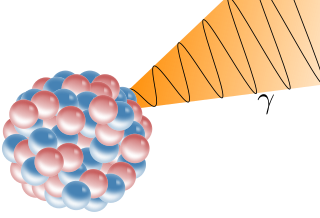
), is a penetrating form of electromagnetic radiation arising from the radioactive decay of atomic nuclei. It consists of the shortest wavelength electromagnetic waves, typically shorter than those of X-rays. With frequencies above 30 exahertz (3×10¹⁹ Hz) and wavelength less than 10 picometer (1×10⁻¹¹ m) gamma ray photons have the highest photon energy of any form of electromagnetic radiation. Paul Villard, a French chemist and physicist, discovered gamma radiation in 1900 while studying radiation emitted by radium. In 1903, Ernest Rutherford named this radiation gamma rays based on their relatively strong penetration of matter; in 1900, he had already named two less penetrating types of decay radiation (discovered by Henri Becquerel) alpha rays and beta rays in ascending order of penetrating power.
Phase-contrast X-ray imaging or phase-sensitive X-ray imaging is a general term for different technical methods that use information concerning changes in the phase of an X-ray beam that passes through an object in order to create its images. Standard X-ray imaging techniques like radiography or computed tomography (CT) rely on a decrease of the X-ray beam's intensity (attenuation) when traversing the sample, which can be measured directly with the assistance of an X-ray detector. However, in phase contrast X-ray imaging, the beam's phase shift caused by the sample is not measured directly, but is transformed into variations in intensity, which then can be recorded by the detector.

In X-ray tubes, the heel effect or, more precisely, the anode heel effect is a variation of the intensity of X-rays emitted by the anode depending on the direction of emission along the anode-cathode axis. X-rays emitted toward the anode are less intense than those emitted perpendicular to the cathode–anode axis or toward the cathode. The effect stems from the absorption of X-ray photons before they leave the anode in which they are produced. The probability of absorption depends on the distance the photons travel within the anode material, which in turn depends on the angle of emission relative to the anode surface.
Spectral imaging is an umbrella term for energy-resolved X-ray imaging in medicine. The technique makes use of the energy dependence of X-ray attenuation to either increase the contrast-to-noise ratio, or to provide quantitative image data and reduce image artefacts by so-called material decomposition. Dual-energy imaging, i.e. imaging at two energy levels, is a special case of spectral imaging and is still the most widely used terminology, but the terms "spectral imaging" and "spectral CT" have been coined to acknowledge the fact that photon-counting detectors have the potential for measurements at a larger number of energy levels.

X-ray emission spectroscopy (XES) is a form of X-ray spectroscopy in which a core electron is excited by an incident x-ray photon and then this excited state decays by emitting an x-ray photon to fill the core hole. The energy of the emitted photon is the energy difference between the involved electronic levels. The analysis of the energy dependence of the emitted photons is the aim of the X-ray emission spectroscopy.
References
- 1 2 3 4 5 6 7 8 9 10 11 12 13 Long1 Smith2 Merrill3, Bruce W.1 Barbara J.2 Vinita3 (2016). Merrill's Atlas of Radiographic Positioning & Procedures (Thirteenth ed.). St. Louis: Elsevier. pp. 52–64. ISBN 978-0-323-26342-9.
{{cite book}}: CS1 maint: date and year (link) CS1 maint: numeric names: authors list (link)