
Low-density lipoprotein (LDL) is one of the five major groups of lipoprotein which transport all fat molecules around the body in the extracellular water. These groups, from least dense to most dense, are chylomicrons, very low-density lipoprotein (VLDL), intermediate-density lipoprotein (IDL), low-density lipoprotein (LDL) and high-density lipoprotein (HDL). LDL delivers fat molecules to cells. LDL is involved in atherosclerosis, a process in which it is oxidized within the walls of arteries.

Atherosclerosis is a pattern of the disease arteriosclerosis in which the wall of the artery develops abnormalities, called lesions. These lesions may lead to narrowing due to the buildup of atheromatous plaque. At onset there are usually no symptoms, but if they develop, symptoms generally begin around middle age. When severe, it can result in coronary artery disease, stroke, peripheral artery disease, or kidney problems, depending on which arteries are affected.

A xanthoma, from Greek ξανθός (xanthós) 'yellow', is a deposition of yellowish cholesterol-rich material that can appear anywhere in the body in various disease states. They are cutaneous manifestations of lipidosis in which lipids accumulate in large foam cells within the skin. They are associated with hyperlipidemias, both primary and secondary types.

Hypercholesterolemia, also called high cholesterol, is the presence of high levels of cholesterol in the blood. It is a form of hyperlipidemia, hyperlipoproteinemia, and dyslipidemia.
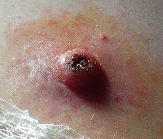
Basal-cell carcinoma (BCC), also known as basal-cell cancer, is the most common type of skin cancer. It often appears as a painless raised area of skin, which may be shiny with small blood vessels running over it. It may also present as a raised area with ulceration. Basal-cell cancer grows slowly and can damage the tissue around it, but it is unlikely to spread to distant areas or result in death.
Hyperlipidemia is abnormally elevated levels of any or all lipids or lipoproteins in the blood. The term hyperlipidemia refers to the laboratory finding itself and is also used as an umbrella term covering any of various acquired or genetic disorders that result in that finding. Hyperlipidemia represents a subset of dyslipidemia and a superset of hypercholesterolemia. Hyperlipidemia is usually chronic and requires ongoing medication to control blood lipid levels.

Arcus senilis (AS), also known as gerontoxon, arcus lipoides, arcus cornae, corneal arcus, arcus adiposus, or arcus cornealis, are rings in the peripheral cornea. It‘s usually caused by cholesterol deposits, so it may be a sign of high cholesterol. It is the most common peripheral corneal opacity, and is usually found in the elderly where it is considered a benign condition. When AS is found in patients less than 50 years old it is termed arcus juvenilis. The finding of arcus juvenilis in combination with hyperlipidemia in younger men represents an increased risk for cardiovascular disease.

Lentigo maligna is where melanocyte cells have become malignant and grow continuously along the stratum basale of the skin, but have not invaded below the epidermis. Lentigo maligna is not the same as lentigo maligna melanoma, as detailed below. It typically progresses very slowly and can remain in a non-invasive form for years.
Keratoacanthoma (KA) is a common low-grade rapidly-growing skin tumour that is believed to originate from the hair follicle and can resemble squamous cell carcinoma.

Sitosterolemia is a rare autosomal recessively inherited lipid metabolic disorder. It is characterized by hyperabsorption and decreased biliary excretion of dietary sterols. Healthy persons absorb only about 5% of dietary plant sterols, but sitosterolemia patients absorb 15% to 60% of ingested sitosterol without excreting much into the bile. The phytosterol campesterol is more readily absorbed than sitosterol.
Familial hypercholesterolemia (FH) is a genetic disorder characterized by high cholesterol levels, specifically very high levels of low-density lipoprotein, in the blood and early cardiovascular disease. The most common mutations diminish the number of functional LDL receptors in the liver. Since the underlying body biochemistry is slightly different in individuals with FH, their high cholesterol levels are less responsive to the kinds of cholesterol control methods which are usually more effective in people without FH. Nevertheless, treatment is usually effective.

Juvenile xanthogranuloma is a form of histiocytosis, classified as "non-Langerhans cell histiocytosis", or more specifically, "type 2".
Verruciform xanthoma is an uncommon benign lesion that has a verruciform (wart-like) appearance, but it may appear polypoid, papillomatous, or sessile. The verruciform was first described by Shafer in 1971 on the oral mucosa. Usually found on the oral mucosa of middle-aged persons, verruciform xanthomas have also been reported on the scrotum and penis of middle-aged to elderly Japanese males. While the most common site is the oral mucosa, lesions that occur elsewhere usually arise on the perineum or on the skin with some predisposing factor, such as lymphedema or an epidermal nevus.
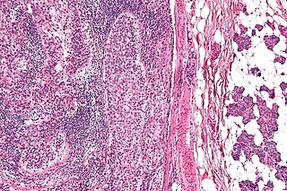
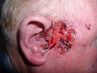
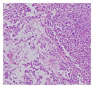
Sebaceous carcinoma, also known as sebaceous gland carcinoma (SGc), sebaceous cell carcinoma, and meibomian gland carcinoma is an uncommon malignant cutaneous tumor. Most are typically about 1.4 cm at presentation. SGc originates from sebaceous glands in the skin and, therefore, may originate anywhere in the body where these glands are found. SGc can be divided into 2 types: periocular and extraocular. The periocular region is rich in sebaceous glands making it a common site of origin. The cause of these lesions in the vast majority of cases is unknown. Occasional cases may be associated with Muir-Torre syndrome. SGc accounts for approximately 0.7% of all skin cancers, and the incidence of SGc is highest in Caucasian, Asian, and Indian populations. Due to the rarity of this tumor and variability in clinical and histological presentation, SGc is often misdiagnosed as an inflammatory condition or a more common neoplasm. SGc is commonly treated with wide local excision or Mohs micrographic surgery, and the relative survival rates at 5 and 10 years are 92.72 and 86.98%, respectively.

A preauricular sinus is a common congenital malformation characterized by a nodule, dent or dimple located anywhere adjacent to the external ear. Frequency of preauricular sinus differs depending the population: 0.1–0.9% in the US, 0.9% in the UK, and 4–10% in Asia and parts of Africa.

Koenen's tumor (KT), also commonly termed periungual angiofibroma, is a subtype of the angiofibromas. Angiofibromas are benign papule, nodule, and/or tumor lesions that are separated into various subtypes based primarily on the characteristic locations of their lesions. KTs are angiofibromas that develop in and under the toenails and/or fingernails. KTs were once considered as the same as another subtype of the angiofibromas viz., acral angiofibromas. While the literature may still sometimes regard KTs as acral angiofibromas, acral angiofibromas are characteristically located in areas close to but not in the toenails and fingernails as well as in the soles of the feet and palms of the hands. KTs are here regarded as distinct from acral angiofibromas.

Familial dysbetalipoproteinemia or type III hyperlipoproteinemia is a condition characterized by increased total cholesterol and triglyceride levels, and decreased HDL levels.
A malignant chondroid syringoma is a very uncommon cutaneous (skin) condition characterised by an adnexal eccrine tumour.

Touton giant cells are a type of multinucleated giant cell seen in lesions with high lipid content such as fat necrosis, xanthoma, and xanthelasma and xanthogranulomas. They are also found in dermatofibroma.

Palisaded encapsulated neuroma (PEN) is a rare, benign cutaneous condition characterized by small, firm, non-pigmented nodules or papules. They typically occur as a solitary (single) lesion near the mucocutaneous junction of the skin of the face, although they can occur elsewhere on the body.
