Amniotic epithelial cell

Last updated

An amniotic epithelial cell is a form of stem cell extracted from the lining of the inner membrane of the placenta. Amniotic epithelial cells start to develop around 8 days post fertilization. These cells are known to have some of the same markers as embryonic stem cells, more specifically, Oct-4 and nanog. These transcription factors are the basis of the pluripotency of stem cells. Amniotic epithelial cells have the ability to develop into any of the three germ layers: endoderm, mesoderm, and ectoderm. They can develop into several organ tissues specific to these germ layers including heart, brain, and liver. The pluripotency of the human amniotic epithelial cells makes them useful in treating and fighting diseases and disorders of the nervous system as well as other tissues of the human body. Artificial heart valves and working tracheas, as well as muscle, fat, bone, heart, neural and liver cells have all been engineered using amniotic stem cells. Tissues obtained from amniotic cell lines show promise for patients with congenital diseases or malformations of the heart, liver, lungs, kidneys, and cerebral tissue.

Contents

Transplantation

Amniotic epithelial cells have shown to be safe and successfully transplanted into hosts that are not related to the donor. One possible reason for this is that amniotic epithelial cells have low antigen levels that inhibit compatibility from a donor to the recipient. This makes the possibility of rejection of the cells by the recipient tissue less likely. Unlike traditional tissue transplants, where the donor and recipient have to be in complete sync as far as blood type and overall compatibility and even still can show tissue rejection, amniotic epithelial cells can be used and show a higher likelihood of tissue acceptance. Also, amniotic epithelial cells are known to promote natural wound healing as well as the inhibition of angiogenesis, which is the basic conversion of a tumor into malignant cancer.

Advantages over embryonic stem cells

In harvesting embryonic stem cells, a human embryo is destroyed. Many anti-abortion individuals associate this act with abortion and consider it immoral. Amniotic epithelial cells are harvested from the placenta, which is commonly discarded after birth. The cells are very easily obtainable without the use of intrusive procedures. Thus their use averts the controversy about embryonic stem cells. There are also large amounts of amniotic epithelial cells found in the placenta and can be found in upwards of 50-100 million cells from one extraction. However, several billion cells are required to use for transplantation in order to treat and fight diseases. Therefore, the cells must be expanded in a lab to have enough cells for transplantation.

Unlike embryonic stem cells, amniotic stem cells have not shown a propensity for developing into teratomas and other cancer-like tumors upon injection into living tissue. Amniotic epithelial cells have not been known to produce cancerous tumors in the host despite the fact that these cells do express genes found in embryonic stem cells that are known to promote tumor formation.

Organs engineered from amniotic epithelial cells obtained from the placenta associated with a particular person's birth would not be rejected by that person; such organs would have the same genotype as that person and thus be fully compatible with that person's immune system.

Expansion

Because there is not a sufficient amount of amniotic epithelial cells extracted from a single term placenta to be use in cellular therapies, the cells must be expanded in a lab. However, some studies have shown that once these cells are duplicated in the lab, some of the pluripotency is lost. Also, these lab grown cells have shown altered gene expression causing a differing phenotype as well as differing antigen levels. This may change their ability to be compatible across non related tissues. Much of the effects of expanding these cells are unknown and scientists are continuing to study ways to cultivate these cells without changing their pluripotent properties. Scientists[ who? ] have found that freezing amniotic epithelial cells causes them to not function as they normally would, which have scientists thinking that the extracellular matrix is the part of the cell that controls its functions.

Cellular therapies

There have been several studies conducted on the potential benefits of using amniotic epithelial cells in various parts of the body. One prospective use of these cells includes cellular therapies aimed at dropping inflammation and scarring. Models have shown that using these cells to reduce such inflammation has shown rewarding affects in the lungs and liver. More specifically, amniotic epithelial cells have been used in the past to treat genetic liver diseases such as ornithine transcarbamylase deficiency, familial hypercholesterolemia, and Crigler–Najjar syndrome. These cells are being looked at by scientists as a new and more efficient way to treat diseases of the liver particularly because of the problem arising from a lack of liver donors. Scientists are also working with genetically modified human amniotic epithelial cells in new experimental procedures and cellular therapies.

Also, amniotic epithelial cells have shown potential for encouraging tissue repair of the recipient. Studies have shown that the use of amniotic epithelial cells in cellular therapies involving spinal cord injuries is promising because of their ability to differentiate into fully operational neurons and can release neurotransmitters. These cells can also be used to treat diseases that affect the central nervous system as well as other neurological disorders.

Amniotic epithelial cells are able to make and release acetylcholine and catecholamine. They also show gene expression for dopamine receptors and transporters. Because of this, they are also studied by scientists who research effects of new drugs on dopamine receptors and transporters as well as the basic functions including dopamine secretion and uptake. Several scientists have concluded using lab rats with Parkinson's disease that these cells, when transplanted, reversed the effects of the disease by replacing those dopamine releasing neurons that had died and prevented other neurons from being destroyed by the disease. Currently, Parkinson's disease is treated with dopamine replacement therapy but is not functional with the late progression of the disease and can't cure the disease. Also, other cells that have been used in the past for transplantation to treat Parkinson's, such as neural stem cells and embryonic stem cells, are either limited or controversial in their retrieval. Similar positive effects have also been shown in studies involving chickens with neurological disorders.

Experiments with lab mice have concluded that amniotic epithelial cells can also differentiate into cells of the pancreas that function to produce insulin and regulate blood sugar levels. This could be a possible cure or treatment for diabetes of both types.

These stem cells can also be used in the lung because they are able to differentiate into cells that produce surfactant, which promotes lung development in unborn fetuses. This could be used as treatment for babies born prematurely that have underdeveloped lung capacities. Another promising use of amniotic epithelial cells is to apply them to the cornea to restore its function in individuals with cornea failure. Studies have shown that when these cells are applied, it decreases the swelling of the ocular plane. Amniotic epithelial cells have also shown positive effects when used to treat severe burn victims by encouraging tissue repair of the recipient as well as treatment for certain autoimmune diseases.

Researchers from the Kyoto Prefectural University of Medicine recently conducted a study in which they transplanted amniotic epithelial cells into the oral cavity to treat oral mucosal defects. Because skin grafts taken from the oral cavity cause defects on the donor site, scientists were looking for a better way of treating these defects without creating defects from transplantation. These scientists used lab rabbits with the oral mucosal defects and transplanted cultured amniotic epithelial cells to the defected areas. The cultured amniotic epithelial cells showed markers found on the oral mucosal cells such as certain keratins. They found that the amniotic epithelial cells differentiation into mucosal-like cells and remained on the mucosal defect and therefore could be a possible mechanism for treating those defects. Scientists have recently discovered that human amniotic epithelial cells are able to produce and release the plasma protein albumin.

Related Research Articles

<span class="mw-page-title-main">Human cloning</span> Creation of a genetically identical copy of a human

Therapeutic cloning would involve cloning cells from a human for use in medicine and transplants. It is an active area of research, but is not in medical practice anywhere in the world, as of 2023. Two common methods of therapeutic cloning that are being researched are somatic-cell nuclear transfer and pluripotent stem cell induction.

<span class="mw-page-title-main">Stem cell</span> Undifferentiated biological cells that can differentiate into specialized cells

In multicellular organisms, stem cells are undifferentiated or partially differentiated cells that can differentiate into various types of cells and proliferate indefinitely to produce more of the same stem cell. They are the earliest type of cell in a cell lineage. They are found in both embryonic and adult organisms, but they have slightly different properties in each. They are usually distinguished from progenitor cells, which cannot divide indefinitely, and precursor or blast cells, which are usually committed to differentiating into one cell type.

<span class="mw-page-title-main">Hematopoietic stem cell transplantation</span> Medical procedure to replace blood or immune stem cells

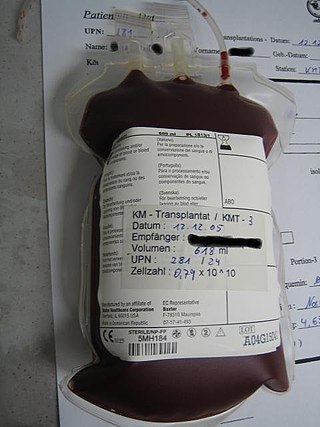
Hematopoietic stem-cell transplantation (HSCT) is the transplantation of multipotent hematopoietic stem cells, usually derived from bone marrow, peripheral blood, or umbilical cord blood in order to replicate inside of a patient and to produce additional normal blood cells. It may be autologous, allogeneic or syngeneic.

<span class="mw-page-title-main">Graft-versus-host disease</span> Medical condition

Graft-versus-host disease (GvHD) is a syndrome, characterized by inflammation in different organs. GvHD is commonly associated with bone marrow transplants and stem cell transplants.

<span class="mw-page-title-main">Embryonic stem cell</span> Type of pluripotent blastocystic stem cell

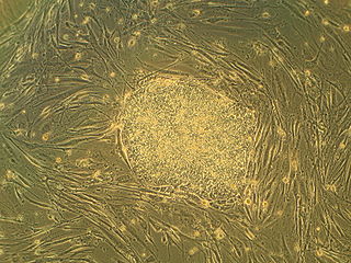
Embryonic stem cells (ESCs) are pluripotent stem cells derived from the inner cell mass of a blastocyst, an early-stage pre-implantation embryo. Human embryos reach the blastocyst stage 4–5 days post fertilization, at which time they consist of 50–150 cells. Isolating the inner cell mass (embryoblast) using immunosurgery results in destruction of the blastocyst, a process which raises ethical issues, including whether or not embryos at the pre-implantation stage have the same moral considerations as embryos in the post-implantation stage of development.

<span class="mw-page-title-main">Regenerative medicine</span> Field of medicine involved in regenerating tissues

Regenerative medicine deals with the "process of replacing, engineering or regenerating human or animal cells, tissues or organs to restore or establish normal function". This field holds the promise of engineering damaged tissues and organs by stimulating the body's own repair mechanisms to functionally heal previously irreparable tissues or organs.

<span class="mw-page-title-main">Adult stem cell</span> Multipotent stem cell in the adult body

Adult stem cells are undifferentiated cells, found throughout the body after development, that multiply by cell division to replenish dying cells and regenerate damaged tissues. Also known as somatic stem cells, they can be found in juvenile, adult animals, and humans, unlike embryonic stem cells.

The stem cell controversy is the consideration of the ethics of research involving the development and use of human embryos. Most commonly, this controversy focuses on embryonic stem cells. Not all stem cell research involves human embryos. For example, adult stem cells, amniotic stem cells, and induced pluripotent stem cells do not involve creating, using, or destroying human embryos, and thus are minimally, if at all, controversial. Many less controversial sources of acquiring stem cells include using cells from the umbilical cord, breast milk, and bone marrow, which are not pluripotent.

Stem-cell therapy is the use of stem cells to treat or prevent a disease or condition. As of 2016, the only established therapy using stem cells is hematopoietic stem cell transplantation. This usually takes the form of a bone-marrow transplantation, but the cells can also be derived from umbilical cord blood. Research is underway to develop various sources for stem cells as well as to apply stem-cell treatments for neurodegenerative diseases and conditions such as diabetes and heart disease.

<span class="mw-page-title-main">Organoid</span> Miniaturized and simplified version of an organ

An organoid is a miniaturized and simplified version of an organ produced in vitro in three dimensions that mimics the key functional, structural and biological complexity of that organ. They are derived from one or a few cells from a tissue, embryonic stem cells or induced pluripotent stem cells, which can self-organize in three-dimensional culture owing to their self-renewal and differentiation capacities. The technique for growing organoids has rapidly improved since the early 2010s, and The Scientist names it as one of the biggest scientific advancements of 2013. Scientists and engineers use organoids to study development and diseases in the laboratory and industry for drug discovery and development, personalized diagnostics and medicine, gene and cell therapies, tissue engineering and regenerative medicine.

<span class="mw-page-title-main">Anthony Atala</span> American bioengineer and urologist

Anthony Atala is an American bioengineer, urologist, and pediatric surgeon. He is the W.H. Boyce professor of urology, the founding director of the Wake Forest Institute for Regenerative Medicine, and the chair of the Department of Urology at Wake Forest School of Medicine in North Carolina. His work focuses on the science of regenerative medicine: "a practice that aims to refurbish diseased or damaged tissue using the body's own healthy cells".

<span class="mw-page-title-main">Induced pluripotent stem cell</span> Pluripotent stem cell generated directly from a somatic cell

Induced pluripotent stem cells are a type of pluripotent stem cell that can be generated directly from a somatic cell. The iPSC technology was pioneered by Shinya Yamanaka and Kazutoshi Takahashi in Kyoto, Japan, who together showed in 2006 that the introduction of four specific genes, collectively known as Yamanaka factors, encoding transcription factors could convert somatic cells into pluripotent stem cells. Shinya Yamanaka was awarded the 2012 Nobel Prize along with Sir John Gurdon "for the discovery that mature cells can be reprogrammed to become pluripotent."

Amniotic stem cells are the mixture of stem cells that can be obtained from the amniotic fluid as well as the amniotic membrane. They can develop into various tissue types including skin, cartilage, cardiac tissue, nerves, muscle, and bone. The cells also have potential medical applications, especially in organ regeneration.

Adult mesenchymal stem cells are being used by researchers in the fields of regenerative medicine and tissue engineering to artificially reconstruct human tissue which has been previously damaged. Mesenchymal stem cells are able to differentiate, or mature from a less specialized cell to a more specialized cell type, to replace damaged tissues in various organs.

Induced stem cells (iSC) are stem cells derived from somatic, reproductive, pluripotent or other cell types by deliberate epigenetic reprogramming. They are classified as either totipotent (iTC), pluripotent (iPSC) or progenitor or unipotent – (iUSC) according to their developmental potential and degree of dedifferentiation. Progenitors are obtained by so-called direct reprogramming or directed differentiation and are also called induced somatic stem cells.

Cord lining, cord tissue, or umbilical cord lining membrane, is the outermost layer of the umbilical cord. As the umbilical cord itself is an extension of the placenta, the umbilical cord lining membrane is an extension of the amniotic membrane covering the placenta. The umbilical cord lining membrane comprises two layers: the amniotic layer and the sub-amniotic layer. The umbilical cord lining membrane is a rich source of two strains of stem cells (CLSCs): epithelial stem cells (CLECs) and mesenchymal stem cells (CLMCs). Discovered by Singapore-based CellResearch Corporation in 2004, this is the best known source for harvesting human stem cells.

Regeneration in humans is the regrowth of lost tissues or organs in response to injury. This is in contrast to wound healing, or partial regeneration, which involves closing up the injury site with some gradation of scar tissue. Some tissues such as skin, the vas deferens, and large organs including the liver can regrow quite readily, while others have been thought to have little or no capacity for regeneration following an injury.

<span class="mw-page-title-main">Limbal stem cell</span>

Limbal stem cells, also known as corneal epithelial stem cells, are unipotent stem cells located in the basal epithelial layer of the corneal limbus. They form the border between the cornea and the sclera. Characteristics of limbal stem cells include a slow turnover rate, high proliferative potential, clonogenicity, expression of stem cell markers, as well as the ability to regenerate the entire corneal epithelium. Limbal stem cell proliferation has the role of maintaining the cornea; for example, by replacing cells that are lost via tears. Additionally, these cells also prevent the conjunctival epithelial cells from migrating onto the surface of the cornea.

Lorenz Studer is a Swiss biologist. He is the founder and director of the Center for Stem Cell Biology at Memorial-Sloan Kettering Cancer Center in New York City. He is a developmental biologist and neuroscientist who is pioneering the generation of midbrain dopamine neurons for transplantation and clinical applications. His expertise in cell engineering spans a wide range of cells/tissues within the nervous system geared toward disease modeling and exploring cell replacement therapy. Currently, he is a member of the Developmental Biology Program and Department of Neurosurgery at Memorial Sloan-Kettering Cancer Center and a Professor of Neuroscience at Weill Cornell Medical College in New York City, NY.

Pregnancy-specific biological substances, which include the placenta, umbilical cord, amniotic fluid, and amniotic membrane are being studied for a number of health uses. For example, Placental-derived stem cells are being studied so they can serve as a potential treatment method for cell therapy. Hepatocyte-like cells (HLC) are generated from differentiated human amniotic epithelial cells (hAEC) that are abundant in the placenta. HLC may replace hepatocytes for hepatocyte transplantation to treat acute or chronic liver damage.

References