Related Research Articles

The basal ganglia are a group of subcortical nuclei, of varied origin, in the brains of vertebrates. In humans, and some primates, there are some differences, mainly in the division of the globus pallidus into an external and internal region, and in the division of the striatum. The basal ganglia are situated at the base of the forebrain and top of the midbrain. Basal ganglia are strongly interconnected with the cerebral cortex, thalamus, and brainstem, as well as several other brain areas. The basal ganglia are associated with a variety of functions, including control of voluntary motor movements, procedural learning, habit learning, conditional learning, eye movements, cognition, and emotion.
In the neuroanatomy of animals, an avian pallium is the dorsal telencephalon of a bird's brain. The subpallium is the ventral telencephalon.

The internal capsule is a white matter structure situated in the inferomedial part of each cerebral hemisphere of the brain. It carries information past the basal ganglia, separating the caudate nucleus and the thalamus from the putamen and the globus pallidus. The internal capsule contains both ascending and descending axons, going to and coming from the cerebral cortex. It also separates the caudate nucleus and the putamen in the dorsal striatum, a brain region involved in motor and reward pathways.

The lateral ventricles are the two largest ventricles of the brain and contain cerebrospinal fluid (CSF). Each cerebral hemisphere contains a lateral ventricle, known as the left or right ventricle, respectively.

The lobes of the brain were originally a purely anatomical classification, but have been shown also to be related to different brain functions. The cerebrum, the largest portion of the human brain, is divided into lobes, but so is the cerebellum. If not specified, the expression "lobes of the brain" refers to the cerebrum.

Brodmann area 24 is part of the anterior cingulate in the human brain.

The septal area, consisting of the lateral septum and medial septum, is an area in the lower, posterior part of the medial surface of the frontal lobe, and refers to the nearby septum pellucidum.
The amygdalofugal pathway is one of the three major efferent pathways of the amygdala, meaning that it is one of the three principal pathways by which fibers leave the amygdala. It leads from the basolateral nucleus and central nucleus of the amygdala. The amygdala is a limbic structure in the medial temporal lobe of the brain. The other main efferent pathways from the amygdala are the stria terminalis and anterior commissure.

The olfactory tract is a bilateral bundle of afferent nerve fibers from the mitral and tufted cells of the olfactory bulb that connects to several target regions in the brain, including the piriform cortex, amygdala, and entorhinal cortex. It is a narrow white band, triangular on coronal section, the apex being directed upward.

The anterior olfactory nucleus is a portion of the forebrain of vertebrates.

HVC is a nucleus in the brain of the songbirds necessary for both the learning and the production of bird song. It is located in the lateral caudal nidopallium and has projections to both the direct and the anterior forebrain pathways.
The isothalamus is a division used by some researchers in describing the thalamus.

In the ventral cochlear nucleus (VCN), auditory nerve fibers enter the brain via the nerve root in the VCN. The ventral cochlear nucleus is divided into the anterior ventral (anteroventral) cochlear nucleus (AVCN) and the posterior ventral (posteroventral) cochlear nucleus (PVCN). In the VCN, auditory nerve fibers bifurcate, the ascending branch innervates the AVCN and the descending branch innervates the PVCN and then continue to the dorsal cochlear nucleus. The orderly innervation by auditory nerve fibers gives the AVCN a tonotopic organization along the dorsoventral axis. Fibers that carry information from the apex of the cochlea that are tuned to low frequencies contact neurons in the ventral part of the AVCN; those that carry information from the base of the cochlea that are tuned to high frequencies contact neurons in the dorsal part of the AVCN. Several populations of neurons populate the AVCN. Bushy cells receive input from auditory nerve fibers through particularly large endings called end bulbs of Held. They contact stellate cells through more conventional boutons.
Vocal learning is the ability to modify acoustic and syntactic sounds, acquire new sounds via imitation, and produce vocalizations. "Vocalizations" in this case refers only to sounds generated by the vocal organ as opposed to by the lips, teeth, and tongue, which require substantially less motor control. A rare trait, vocal learning is a critical substrate for spoken language and has only been detected in eight animal groups despite the wide array of vocalizing species; these include humans, bats, cetaceans, pinnipeds, elephants, and three distantly related bird groups including songbirds, parrots, and hummingbirds. Vocal learning is distinct from auditory learning, or the ability to form memories of sounds heard, a relatively common trait which is present in all vertebrates tested. For example, dogs can be trained to understand the word "sit" even though the human word is not in its innate auditory repertoire. However, the dog cannot imitate and produce the word "sit" itself as vocal learners can.

In neuroanatomy, pallium refers to the layers of grey and white matter that cover the upper surface of the cerebrum in vertebrates. The non-pallial part of the telencephalon builds the subpallium. In basal vertebrates the pallium is a relatively simple three-layered structure, encompassing 3–4 histogenetically distinct domains, plus the olfactory bulb.
The interpeduncular nucleus (IPN) is an unpaired, ovoid cell group at the base of the midbrain tegmentum. It is located in the mesencephalon below the interpeduncular fossa. As the name suggests, the interpeduncular nucleus lies in between the cerebral peduncles.
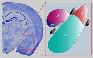
The central nucleus of the amygdala is a nucleus within the amygdala. It "serves as the major output nucleus of the amygdala and participates in receiving and processing pain information."
The dorsal tegmental nucleus (DTN), also known as dorsal tegmental nucleus of Gudden (DTg), is a group of neurons located in the brain stem, which are involved in spatial navigation and orientation.
References
- ↑ "Avian Brain Nomenclature History Table". Archived from the original on 2008-05-13. Retrieved 2007-12-16.
- ↑ "New Terminology for the Archistriatum". Archived from the original on 2008-05-13. Retrieved 2007-12-16.
- ↑ Reiner A, Perkel DJ, Bruce LL, et al. (May 2004). "Revised nomenclature for avian telencephalon and some related brainstem nuclei". J. Comp. Neurol. 473 (3): 377–414. doi:10.1002/cne.20118. PMC 2518311 . PMID 15116397.