Related Research Articles

The breast is one of two prominences located on the upper ventral region of a primate's torso. Both females and males develop breasts from the same embryological tissues.

Gastroenterology is the branch of medicine focused on the digestive system and its disorders. The digestive system consists of the gastrointestinal tract, sometimes referred to as the GI tract, which includes the esophagus, stomach, small intestine and large intestine as well as the accessory organs of digestion which include the pancreas, gallbladder, and liver. The digestive system functions to move material through the GI tract via peristalsis, break down that material via digestion, absorb nutrients for use throughout the body, and remove waste from the body via defecation. Physicians who specialize in the medical specialty of gastroenterology are called gastroenterologists or sometimes GI doctors. Some of the most common conditions managed by gastroenterologists include gastroesophageal reflux disease, gastrointestinal bleeding, irritable bowel syndrome, inflammatory bowel disease (IBD) which includes Crohn's disease and ulcerative colitis, peptic ulcer disease, gallbladder and biliary tract disease, hepatitis, pancreatitis, colitis, colon polyps and cancer, nutritional problems, and many more.

The nipple is a raised region of tissue on the surface of the breast from which, in females, milk leaves the breast through the lactiferous ducts to feed an infant. The milk can flow through the nipple passively or it can be ejected by smooth muscle contractions that occur along with the ductal system. Male mammals also have nipples but without the same level of function, and often surrounded by body hair.
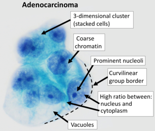
Adenocarcinoma is a type of cancerous tumor that can occur in several parts of the body. It is defined as neoplasia of epithelial tissue that has glandular origin, glandular characteristics, or both. Adenocarcinomas are part of the larger grouping of carcinomas, but are also sometimes called by more precise terms omitting the word, where these exist. Thus invasive ductal carcinoma, the most common form of breast cancer, is adenocarcinoma but does not use the term in its name—however, esophageal adenocarcinoma does to distinguish it from the other common type of esophageal cancer, esophageal squamous cell carcinoma. Several of the most common forms of cancer are adenocarcinomas, and the various sorts of adenocarcinoma vary greatly in all their aspects, so that few useful generalizations can be made about them.

A mammary gland is an exocrine gland in humans and other mammals that produces milk to feed young offspring. Mammals get their name from the Latin word mamma, "breast". The mammary glands are arranged in organs such as the breasts in primates, the udder in ruminants, and the dugs of other animals. Lactorrhea, the occasional production of milk by the glands, can occur in any mammal, but in most mammals, lactation, the production of enough milk for nursing, occurs only in phenotypic females who have gestated in recent months or years. It is directed by hormonal guidance from sex steroids. In a few mammalian species, male lactation can occur. With humans, male lactation can occur only under specific circumstances.

Microcalcifications are tiny deposits of calcium salts that are too small to be felt but can be detected by imaging.
The tail of Spence has historically been described as an extension of the tissue of the upper outer quadrant of the breast traveling into the axilla. The "axillary tail" has been reported to pass into the axilla through an opening in the deep fascia called foramen of Langer. The "tail of Spence" was named after the Scottish surgeon James Spence, who served as a President of the Royal College of Surgeons in Edinburgh in the latter half of the 19th Century.
Atypical hyperplasia is a benign (noncancerous) cellular hyperplasia in which cells show some atypia. In this condition, cells look abnormal under a microscope and are increased in number.
Benign proliferative breast disease is a group of noncancerous conditions that may increase the risk of developing breast cancer. Examples include atypical ductal hyperplasia, atypical lobular hyperplasia, and intraductal papillomas.
Cholangiosarcoma is a tumor of the connective tissues of the bile ducts.
Stereotactic biopsy, also known as stereotactic core biopsy, is a biopsy procedure that uses a computer and imaging performed in at least two planes to localize a target lesion in three-dimensional space and guide the removal of tissue for examination by a pathologist under a microscope. Stereotactic core biopsy makes use of the underlying principle of parallax to determine the depth or "Z-dimension" of the target lesion.

Medullary breast carcinoma is a rare type of breast cancer that is characterized as a relatively circumscribed tumor with pushing, rather than infiltrating, margins. It is histologically characterized as poorly differentiated cells with abundant cytoplasm and pleomorphic high grade vesicular nuclei. It involves lymphocytic infiltration in and around the tumor and can appear to be brown in appearance with necrosis and hemorrhage. Prognosis is measured through staging but can often be treated successfully and has a better prognosis than other infiltrating breast carcinomas.
Metaplastic carcinoma, otherwise known as metaplastic carcinoma of the breast (MCB), is a heterogeneous group of cancers that exhibit varied patterns of metaplasia and differentiation along multiple cell lines. This rare and aggressive form of breast cancer is characterized as being composed of a mixed group of neoplasms containing both glandular and non-glandular patterns with epithelial and/or mesenchymal components. It accounts for fewer than 1% of all breast cancer diagnoses. It is most closely associated with invasive ductal carcinoma of no special type. (IDC), and shares similar treatment approaches. Relative to IDC, MCB generally has higher histological grade and larger tumor size at time of diagnosis, with a lower incidence of axillary lymph node involvement. MCB tumors are typically estrogen receptor (ER), progesterone receptor (PR), and human epidermal growth factor-2 (HER-2) negative, meaning hormone therapy is generally not an effective treatment option, which correlates to a relatively poor prognosis. MCB was first recognized as a distinct pathological entity in 2000 by the World Health Organization.

Salivary gland tumours, also known as mucous gland adenomas or neoplasms, are tumours that form in the tissues of salivary glands. The salivary glands are classified as major or minor. The major salivary glands consist of the parotid, submandibular, and sublingual glands. The minor salivary glands consist of 800 to 1000 small mucus-secreting glands located throughout the lining of the oral cavity. Patients with these types of tumours may be asymptomatic.

Molecular breast imaging (MBI), also known as scintimammography, is a type of breast imaging test that is used to detect cancer cells in breast tissue of individuals who have had abnormal mammograms, especially for those who have dense breast tissue, post-operative scar tissue or breast implants.
Fatty-replaced breast tissue is a term used in mammography that refers to the replacement of breast tissue with fatty tissue. This commonly occurs as a woman ages.
Needle-localized biopsy is a procedure that uses very thin needles or guide wires to mark the location of an abnormal area of tissue so it can be surgically sampled. An imaging device such as an ultrasound probe is used to place the wire in or around the abnormal area. Needle localization is used when the doctor cannot feel the mass of abnormal tissue.
An open biopsy is a procedure in which a surgical incision (cut) is made through the skin to expose and remove tissues. The biopsy tissue is examined under a microscope by a pathologist. An open biopsy may be done in the doctor's office or hospital, and may use local anesthesia or general anesthesia. A lumpectomy to remove a breast tumor is a type of open biopsy.
Comedocarcinoma is a kind of breast cancer that demonstrates comedonecrosis, which is the central necrosis of cancer cells within involved ducts. Comedocarcinomas are usually non-infiltrating and intraductal tumors, characterized as a comedo-type, high-grade ductal carcinoma in situ (DCIS). However, there have been accounts of comedocarcinoma which has then diversified into other cell types and developed into infiltrating (invasive) ductal carcinoma. Recurrence and survival rates differ for invasive breast cancer which has originated as comedocarcinoma compared with other types of cancer cells.
A fungating lesion is a skin lesion that fungates, that is, becomes like a fungus in its appearance or growth rate. It is marked by ulcerations and necrosis and usually presents a foul odor. This kind of lesion may occur in many types of cancer, including breast cancer, melanoma, and squamous cell carcinoma, and especially in advanced disease. The characteristic malodorous smell is caused by dimethyl trisulfide. It is usually not a fungal infection but rather a neoplastic growth with necrosing portions.
References
- Breast duct endoscopy entry in the public domain NCI Dictionary of Cancer Terms
![]() This article incorporates public domain material from Dictionary of Cancer Terms. U.S. National Cancer Institute.
This article incorporates public domain material from Dictionary of Cancer Terms. U.S. National Cancer Institute.
| | This oncology article is a stub. You can help Wikipedia by expanding it. |
| | This article related to medical imaging is a stub. You can help Wikipedia by expanding it. |