This page is based on this
Wikipedia article Text is available under the
CC BY-SA 4.0 license; additional terms may apply.
Images, videos and audio are available under their respective licenses.

The mandibular nerve (V3) is the largest of the three divisions of the trigeminal nerve, the fifth cranial nerve (CN V).

The buccinator (
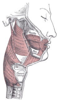
) is a thin quadrilateral muscle occupying the interval between the maxilla and the mandible at the side of the face. It forms the anterior part of the cheek or the lateral wall of the oral cavity.

The zygomaticus major is a muscle of the human body. It is a muscle of facial expression which draws the angle of the mouth superiorly and posteriorly to allow one to smile. Like all muscles of facial expression, the zygomatic major is innervated by the facial nerve, more specifically, the buccal and zygomatic branches of the facial nerve.

The levator anguli oris (caninus) is a facial muscle of the mouth arising from the canine fossa, immediately below the infraorbital foramen. It elevates angle of mouth medially. Its fibers are inserted into the angle of the mouth, intermingling with those of the zygomaticus, triangularis, and orbicularis oris. Specifically, the levator anguli oris is innervated by the buccal branches of the facial nerve.
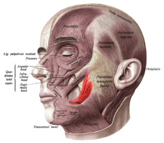
The risorius is a muscle of facial expression which arises in the fascia over the parotid gland and, passing horizontally forward, superficial to the platysma, inserts onto the skin at the angle of the mouth. It is a narrow bundle of fibers, broadest at its origin, but varies much in its size and form.
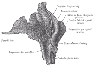
The parotid duct or Stensen duct is a duct and the route that saliva takes from the major salivary gland, the parotid gland into the mouth.
The oral mucosa is the mucous membrane lining the inside of the mouth and consists of stratified squamous epithelium termed oral epithelium and an underlying connective tissue termed lamina propria. The oral cavity has sometimes been described as a mirror that reflects the health of the individual. Changes indicative of disease are seen as alterations in the oral mucosa lining the mouth, which can reveal systemic conditions, such as diabetes or vitamin deficiency, or the local effects of chronic tobacco or alcohol use.
The oral mucosa tends to heal faster and with less scar formation compared to the skin. The underlying mechanism remains unknown but research suggest that extracellular vesicles might be involved.

The buccal nerve is a nerve in the face. It is a branch of the mandibular nerve and transmits sensory information from skin over the buccal membrane and from the second and third molar teeth. Not to be confused with the buccal branch of the facial nerve which transmits motor information to the buccinator muscle.

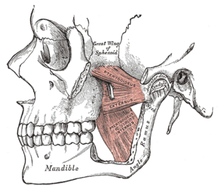
The infratemporal fossa is an irregularly shaped cavity, situated below and medial to the zygomatic arch. It is not fully enclosed by bone in all directions, and it contains superficial muscles that are visible during dissection after removing skin and fascia: namely, the lower part of the temporalis muscle, the lateral pterygoid, and the medial pterygoid.

The buccal branches of the facial nerve, are of larger size than the rest of the branches, pass horizontally forward to be distributed below the orbit and around the mouth.
A cutaneous nerve is a nerve that provides nerve supply to the skin.
The labial glands are minor salivary glands situated between the mucous membrane and the orbicularis oris around the orifice of the mouth.
The parotid plexus or pes anserinus or plexus parotideus is the branch point of the facial nerve (extratemporal) after it leaves the stylomastoid foramen. This division takes place within the parotid gland.
Inferior alveolar nerve block is a nerve block technique which induces anesthesia (numbness) in the areas of the mouth and face innervated by one of the inferior alveolar nerves which are paired on the left and right side. These areas are the skin and mucous membranes of the lower lip, the skin of the chin, the lower teeth and the labial gingiva of the anterior teeth, all unilaterally to the midline of the side on which the block is administered. However, depending on technique, the long buccal nerve may not be anesthetized by an IANB and therefore an area of buccal gingiva adjacent to the lower posterior teeth will retain normal sensation unless that nerve is anesthetized separately, via a (long) buccal nerve block. The inferior alveolar nerve is a branch of the mandibular nerve, the third division of the trigeminal nerve. This procedure attempts to anaesthetise the inferior alveolar nerve prior to it entering the mandibular foramen on the medial surface of the mandibular ramus.

Buccal fat pad extraction is a plastic surgery procedure for reducing prominent cheeks, by the proportional removal of buccal fat-pad tissue.

In human anatomy, the mouth is the first portion of the alimentary canal that receives food and produces saliva. The oral mucosa is the mucous membrane epithelium lining the inside of the mouth.
The pterygomandibular space is a fascial space of the head and neck. It is a potential space in the head and is paired on each side. It is located between the medial pterygoid muscle and the medial surface of the ramus of the mandible. The pterygomandibular space is one of the four compartments of the masticator space.

The canine space, is a fascial space of the head and neck. It is a thin potential space on the face, and is paired on either side. It is located between the levator anguli oris muscle inferiorly and the levator labii superioris muscle superiorly. The term is derived from the fact that the space is in the region of the canine fossa, and that infections originating from the maxillary canine tooth may spread to involve the space. Infra-orbital is derived from infra- meaning below and orbit which refers to the eye socket.