This page is based on this
Wikipedia article Text is available under the
CC BY-SA 4.0 license; additional terms may apply.
Images, videos and audio are available under their respective licenses.

The circle of Willis is a circulatory anastomosis that supplies blood to the brain and surrounding structures. It is named after Thomas Willis (1621–1675), an English physician.

The internal carotid artery is a major paired artery, one on each side of the head and neck, in human anatomy. They arise from the common carotid arteries where these bifurcate into the internal and external carotid arteries at cervical vertebral level 3 or 4; the internal carotid artery supplies the brain, while the external carotid nourishes other portions of the head, such as face, scalp, skull, and meninges.

The choroid, also known as the choroidea or choroid coat, is the vascular layer of the eye, containing connective tissues, and lying between the retina and the sclera. The human choroid is thickest at the far extreme rear of the eye, while in the outlying areas it narrows to 0.1 mm. The choroid provides oxygen and nourishment to the outer layers of the retina. Along with the ciliary body and iris, the choroid forms the uveal tract.

The internal capsule is a white matter structure situated in the inferomedial part of each cerebral hemisphere of the brain. It carries information past the basal ganglia, separating the caudate nucleus and the thalamus from the putamen and the globus pallidus. The internal capsule contains both ascending and descending axons, going to and coming from the cerebral cortex.

The middle meningeal artery is typically the third branch of the first part of the maxillary artery, one of the two terminal branches of the external carotid artery. After branching off the maxillary artery in the infratemporal fossa, it runs through the foramen spinosum to supply the dura mater and the calvaria. The middle meningeal artery is the largest of the three (paired) arteries that supply the meninges, the others being the anterior meningeal artery and the posterior meningeal artery.

The anterior cerebral artery (ACA) is one of a pair of arteries on the brain that supplies oxygenated blood to most midline portions of the frontal lobes and superior medial parietal lobes. The two anterior cerebral arteries arise from the internal carotid artery and are part of the circle of Willis. The left and right anterior cerebral arteries are connected by the anterior communicating artery.

The ophthalmic artery (OA) is the first branch of the internal carotid artery distal to the cavernous sinus. Branches of the OA supply all the structures in the orbit as well as some structures in the nose, face and meninges. Occlusion of the OA or its branches can produce sight-threatening conditions.

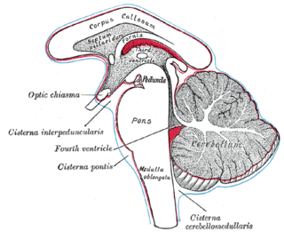
The subarachnoid cisterns are spaces formed by openings in the subarachnoid space, an anatomic space in the meninges of the brain. The space separates two of the meninges, the arachnoid mater and the pia mater. These cisterns are filled with cerebrospinal fluid.

The tela choroidea is a region of meningeal pia mater and underlying ependyma that gives rise to the choroid plexus in each of the brain’s four ventricles. Tela is Latin for woven and is used to describe a web-like membrane or layer. The tela choroidea is a very thin part of the loose connective tissue of pia mater that overlies and closely adheres to the ependyma with no intervening tissue. It has a rich blood supply. The ependyma and vascular pia mater that make up the tela choroidea form regions of minute projections known as a choroid plexus that projects into each ventricle. The choroid plexus produces the cerebrospinal fluid of the ventricular system. The tela choroidea in the ventricles forms from different parts of the roof plate in the development of the embryo.

The princeps pollicis artery, or principal artery of the thumb, arises from the radial artery just as it turns medially towards the deep part of the hand; it descends between the first dorsal interosseous muscle and the oblique head of the adductor pollicis, along the medial side of the first metacarpal bone to the base of the proximal phalanx, where it lies beneath the tendon of the flexor pollicis longus muscle and divides into two branches.
Communicans is a Latin word meaning "communicating". It is most commonly used in medical or biological terminology.
Communicating artery may refer to:
Hemihypesthesia is a reduction in sensitivity on one side of the body. A person with this condition may not be able to perceive being lightly touched on one side, but has normal function on the other side of the body.

The collicular artery or quadrigeminal artery arises from the posterior cerebral artery. This small artery supplies portions of the midbrain, especially the superior colliculus, inferior colliculus, and tectum.
The plexal point is the point at which the anterior choroidal artery enters into the lateral ventricle of the brain at the choroid fissure. The choroid fissure, in this sense, is the narrow cleft along the medial wall of the lateral ventricle, where the choroid plexus is attached at the margins. Not to be confused with the choroidal fissure of the eye.
Cecal artery, caecal artery or arteria caecalis can refer to:
Tympanic artery can refer to:


