Related Research Articles
Tendinopathy is a type of tendon disorder that results in pain, swelling, and impaired function. The pain is typically worse with movement. It most commonly occurs around the shoulder, elbow, wrist, hip, knee, or ankle.

Trigger finger, also known as stenosing tenosynovitis, is a disorder characterized by catching or locking of the involved finger in full or near full flexion, typically with force. There may be tenderness in the palm of the hand near the last skin crease. The name "trigger finger" may refer to the motion of "catching" like a trigger on a gun. The ring finger and thumb are most commonly affected.

A sprain is a soft tissue injury of the ligaments within a joint, often caused by a sudden movement abruptly forcing the joint to exceed its functional range of motion. Ligaments are tough, inelastic fibers made of collagen that connect two or more bones to form a joint and are important for joint stability and proprioception, which is the body's sense of limb position and movement. Sprains may be mild, moderate, or severe, with the latter two classes involving some degree of tearing of the ligament. Sprains can occur at any joint but most commonly occur in the ankle, knee, or wrist. An equivalent injury to a muscle or tendon is known as a strain.

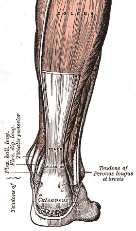
Achilles tendinitis, also known as achilles tendinopathy, occurs when the Achilles tendon, found at the back of the ankle, becomes sore. Achilles tendinopathy is accompanied by alterations in the tendon's structure and mechanical properties. The most common symptoms are pain and swelling around the affected tendon. The pain is typically worse at the start of exercise and decreases thereafter. Stiffness of the ankle may also be present. Onset is generally gradual.
A soft tissue injury is the damage of muscles, ligaments and tendons throughout the body. Common soft tissue injuries usually occur from a sprain, strain, a one-off blow resulting in a contusion or overuse of a particular part of the body. Soft tissue injuries can result in pain, swelling, bruising and loss of function.
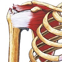
Rotator cuff tendinopathy is a process of senescence. The pathophysiology is mucoid degeneration. Most people develop rotator cuff tendinopathy within their lifetime.
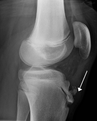
Osgood–Schlatter disease (OSD) is inflammation of the patellar ligament at the tibial tuberosity (apophysitis). It is characterized by a painful bump just below the knee that is worse with activity and better with rest. Episodes of pain typically last a few weeks to months. One or both knees may be affected and flares may recur.

Anterior cruciate ligament reconstruction is a surgical tissue graft replacement of the anterior cruciate ligament, located in the knee, to restore its function after an injury. The torn ligament can either be removed from the knee, or preserved before reconstruction through an arthroscopic procedure. ACL repair is also a surgical option. This involves repairing the ACL by re-attaching it, instead of performing a reconstruction. Theoretical advantages of repair include faster recovery and a lack of donor site morbidity, but randomised controlled trials and long-term data regarding re-rupture rates using contemporary surgical techniques are lacking.

Cruciate ligaments are pairs of ligaments arranged like a letter X. They occur in several joints of the body, such as the knee joint, wrist joint and the atlanto-axial joint. In a fashion similar to the cords in a toy Jacob's ladder, the crossed ligaments stabilize the joint while allowing a very large range of motion.
A sprained ankle is an injury where sprain occurs on one or more ligaments of the ankle. It is the most common injury to occur in ball sports, such as basketball, volleyball, football, and racquet sports.
An anterior cruciate ligament injury occurs when the anterior cruciate ligament (ACL) is either stretched, partially torn, or completely torn. The most common injury is a complete tear. Symptoms include pain, an audible cracking sound during injury, instability of the knee, and joint swelling. Swelling generally appears within a couple of hours. In approximately 50% of cases, other structures of the knee such as surrounding ligaments, cartilage, or meniscus are damaged.

Jammed finger is a colloquialism referring to a variety of injuries to the joints of the fingers, resulting from axial loading beyond that which the ligaments can withstand. Common parts of the finger susceptible to this type of injury are ligaments, joints, and bones. The severity of the damage to the finger increases with the magnitude of the force exerted by the external object on the fingertip. Toes may become jammed as well, with similar results.

A tear of a meniscus is a rupturing of one or more of the fibrocartilage strips in the knee called menisci. When doctors and patients refer to "torn cartilage" in the knee, they actually may be referring to an injury to a meniscus at the top of one of the tibiae. Menisci can be torn during innocuous activities such as walking or squatting. They can also be torn by traumatic force encountered in sports or other forms of physical exertion. The traumatic action is most often a twisting movement at the knee while the leg is bent. In older adults, the meniscus can be damaged following prolonged 'wear and tear'. Especially acute injuries can lead to displaced tears which can cause mechanical symptoms such as clicking, catching, or locking during motion of the joint. The joint will be in pain when in use, but when there is no load, the pain goes away.
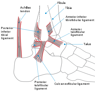
A high ankle sprain, also known as a syndesmotic ankle sprain (SAS), is a sprain of the syndesmotic ligaments that connect the tibia and fibula in the lower leg, thereby creating a mortise and tenon joint for the ankle. High ankle sprains are described as high because they are located above the ankle. They comprise approximately 15% of all ankle sprains. Unlike the common lateral ankle sprains, when ligaments around the ankle are injured through an inward twisting, high ankle sprains are caused when the lower leg and foot externally rotates.

The elbow is the region between the upper arm and the forearm that surrounds the elbow joint. The elbow includes prominent landmarks such as the olecranon, the cubital fossa, and the lateral and the medial epicondyles of the humerus. The elbow joint is a hinge joint between the arm and the forearm; more specifically between the humerus in the upper arm and the radius and ulna in the forearm which allows the forearm and hand to be moved towards and away from the body. The term elbow is specifically used for humans and other primates, and in other vertebrates forelimb plus joint is used.
Jersey finger, also known as rugby finger, is a finger-related tendon injury that is common in sport and can result in permanent loss of flexion of the end of the finger if not surgically repaired. The injury is common when one player grabs another's jersey with the tips of one or more fingers while that player is pulling or running away. It is the most common closed flexor tendon injury and occurs in the ring finger in 75% of cases.

Injuries to the arm, forearm or wrist area can lead to various nerve disorders. One such disorder is median nerve palsy. The median nerve controls the majority of the muscles in the forearm. It controls abduction of the thumb, flexion of hand at wrist, flexion of digital phalanx of the fingers, is the sensory nerve for the first three fingers, etc. Because of this major role of the median nerve, it is also called the eye of the hand. If the median nerve is damaged, the ability to abduct and oppose the thumb may be lost due to paralysis of the thenar muscles. Various other symptoms can occur which may be repaired through surgery and tendon transfers. Tendon transfers have been very successful in restoring motor function and improving functional outcomes in patients with median nerve palsy.
Injuries in rock climbing may occur due to falls, or due to overuse. Injuries due to falls are relatively uncommon; the vast majority of injuries result from overuse, most often occurring in the fingers, elbows, and shoulders. Such injuries are often no worse than torn calluses, cuts, burns and bruises. However, overuse symptoms, if ignored, may lead to permanent damage.
Medial knee injuries are the most common type of knee injury. The medial ligament complex of the knee consists of:

A biceps tendon rupture or bicep tear is a complete or partial rupture of a tendon of the biceps brachii muscle. It can affect any of the three biceps brachii tendons - the proximal tendon of the short head of the muscle belly, the proximal tendon of the long head of the muscle belly, or the distal tendon. The characteristic finding of a biceps tendon rupture is the Popeye sign. Patients often report an audible pop at the time of injury as well as pain, bruising, and swelling. Provocative physical exam maneuvers to assess for a rupture include Ludington's test, Hook test, and the Ruland biceps squeeze test. Treatment and prognosis are highly dependent on the site of the injury described in further detail below.
References
- 1 2 Bollen, S R (1 December 1988). "Soft tissue injury in extreme rock climbers". British Journal of Sports Medicine. 22 (4): 145–147. doi:10.1136/bjsm.22.4.145. PMC 1478743 . PMID 3228682.
- ↑ Preston, Dayton. "Rock Climbing Reaching New Heights". Hughston health alert. Retrieved 12 January 2011.
- 1 2 Hörst, Eric J (2008). "Finger Tendon Pulley Injury". Nicros. Archived from the original on September 30, 2011. Retrieved 12 January 2011.
- ↑ "Climber's Finger: The Pulley Tear". Gripped Magazine. 2020-07-30. Retrieved 2022-01-05.
- ↑ Crowley, Timothy P. (June 2012). "The Flexor Tendon Pulley System and Rock Climbing". Journal of Hand and Microsurgery. 4 (1): 25–29. doi:10.1007/s12593-012-0061-3. PMC 3371120 . PMID 23730085.
- ↑ Roseborrough, Aimee; Roseborrough, Kyle (2009). "Fingers and Pulleys" . Retrieved 12 January 2011.
- ↑ Schöffl, Volker; Schöffl, I. (2007). "Finger pain in rock climbers: reaching the right differential diagnosis and therapy". J Sports Med Phys Fitness. 47 (1): 70–78. PMID 17369801. ProQuest 202716942.
- ↑ Larsson, Robin; Nordeman, Lena; Blomdahl, Christina (2022-08-01). "To tape or not to tape: annular ligament (pulley) injuries in rock climbers—a systematic review". BMC Sports Science, Medicine and Rehabilitation. 14 (1): 148. doi: 10.1186/s13102-022-00539-6 . ISSN 2052-1847. PMC 9344739 . PMID 35915476.