Related Research Articles

The pleural cavity, pleural space, or intrapleural space is the potential space between the pleurae of the pleural sac that surrounds each lung. A small amount of serous pleural fluid is maintained in the pleural cavity to enable lubrication between the membranes, and also to create a pressure gradient.

The respiratory tract is the subdivision of the respiratory system involved with the process of conducting air to the alveoli for the purposes of gas exchange in mammals. The respiratory tract is lined with respiratory epithelium as respiratory mucosa.

Pleurisy, also known as pleuritis, is inflammation of the membranes that surround the lungs and line the chest cavity (pleurae). This can result in a sharp chest pain while breathing. Occasionally the pain may be a constant dull ache. Other symptoms may include shortness of breath, cough, fever, or weight loss, depending on the underlying cause. Pleurisy can be caused by a variety of conditions, including viral or bacterial infections, autoimmune disorders, and pulmonary embolism.

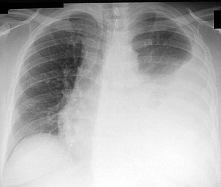
A pleural effusion is accumulation of excessive fluid in the pleural space, the potential space that surrounds each lung. Under normal conditions, pleural fluid is secreted by the parietal pleural capillaries at a rate of 0.6 millilitre per kilogram weight per hour, and is cleared by lymphatic absorption leaving behind only 5–15 millilitres of fluid, which helps to maintain a functional vacuum between the parietal and visceral pleurae. Excess fluid within the pleural space can impair inspiration by upsetting the functional vacuum and hydrostatically increasing the resistance against lung expansion, resulting in a fully or partially collapsed lung.
Radiology (X-rays) is used in the diagnosis of tuberculosis. Abnormalities on chest radiographs may be suggestive of, but are never diagnostic of TB, but can be used to rule out pulmonary TB.

A hemothorax is an accumulation of blood within the pleural cavity. The symptoms of a hemothorax may include chest pain and difficulty breathing, while the clinical signs may include reduced breath sounds on the affected side and a rapid heart rate. Hemothoraces are usually caused by an injury, but they may occur spontaneously due to cancer invading the pleural cavity, as a result of a blood clotting disorder, as an unusual manifestation of endometriosis, in response to pneumothorax, or rarely in association with other conditions.
Thoracentesis, also known as thoracocentesis, pleural tap, needle thoracostomy, or needle decompression, is an invasive medical procedure to remove fluid or air from the pleural space for diagnostic or therapeutic purposes. A cannula, or hollow needle, is carefully introduced into the thorax, generally after administration of local anesthesia. The procedure was first performed by Morrill Wyman in 1850 and then described by Henry Ingersoll Bowditch in 1852.

Respiratory diseases, or lung diseases, are pathological conditions affecting the organs and tissues that make gas exchange difficult in air-breathing animals. They include conditions of the respiratory tract including the trachea, bronchi, bronchioles, alveoli, pleurae, pleural cavity, the nerves and muscles of respiration. Respiratory diseases range from mild and self-limiting, such as the common cold, influenza, and pharyngitis to life-threatening diseases such as bacterial pneumonia, pulmonary embolism, tuberculosis, acute asthma, lung cancer, and severe acute respiratory syndromes, such as COVID-19. Respiratory diseases can be classified in many different ways, including by the organ or tissue involved, by the type and pattern of associated signs and symptoms, or by the cause of the disease.
Hemopneumothorax, or haemopneumothorax, is the condition of having both air (pneumothorax) and blood (hemothorax) in the chest cavity. A hemothorax, pneumothorax, or the combination of both can occur due to an injury to the lung or chest.
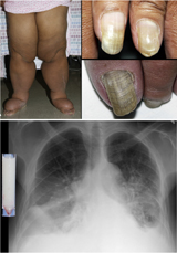
Yellow nail syndrome, also known as "primary lymphedema associated with yellow nails and pleural effusion", is a very rare medical syndrome that includes pleural effusions, lymphedema and yellow dystrophic nails. Approximately 40% will also have bronchiectasis. It is also associated with chronic sinusitis and persistent coughing. It usually affects adults.

Respiratory sounds, also known as lung sounds or breath sounds, are the specific sounds generated by the movement of air through the respiratory system. These may be easily audible or identified through auscultation of the respiratory system through the lung fields with a stethoscope as well as from the spectral characteristics of lung sounds. These include normal breath sounds and added sounds such as crackles, wheezes, pleural friction rubs, stertor, and stridor.
Fremitus is a vibration transmitted through the body. In common medical usage, it usually refers to assessment of the lungs by either the vibration intensity felt on the chest wall and/or heard by a stethoscope on the chest wall with certain spoken words, although there are several other types.

The costodiaphragmatic recess, also called the costophrenic recess or phrenicocostal sinus, is the posterolateral fringe of the pleural space, a potential space around the lung inside the pleural cavity. It is located at the acutely angled junction ("reflection") between the costal and diaphragmatic parietal pleurae, and is interpreted two-dimensionally on plain X-rays as the costophrenic angle. It measures approximately 5 cm (2.0 in) vertically and extends from the eighth to the tenth rib along the mid-axillary line.
Whispered pectoriloquy refers to an increased loudness of whispering noted during auscultation with a stethoscope on the lung fields on a patient's torso.

Subcutaneous emphysema occurs when gas or air accumulates and seeps under the skin, where normally no gas should be present. Subcutaneous refers to the subcutaneous tissue, and emphysema refers to trapped air pockets. Since the air generally comes from the chest cavity, subcutaneous emphysema usually occurs around the upper torso, such as on the chest, neck, face, axillae and arms, where it is able to travel with little resistance along the loose connective tissue within the superficial fascia. Subcutaneous emphysema has a characteristic crackling-feel to the touch, a sensation that has been described as similar to touching warm Rice Krispies. This sensation of air under the skin is known as subcutaneous crepitation, a form of crepitus.
Pleural disease occurs in the pleural space, which is the thin fluid-filled area in between the two pulmonary pleurae in the human body. There are several disorders and complications that can occur within the pleural area, and the surrounding tissues in the lung.

An empyema is a collection or gathering of pus within a naturally existing anatomical cavity. The term is most commonly used to refer to pleural empyema, which is empyema of the pleural cavity. It differs from an abscess, which is a collection of pus in a newly formed cavity. Empyema most commonly occurs as a complication of pneumonia but can also result from other infections or conditions that lead to the collection of infected fluid in a body cavity.

Fibrothorax is a medical condition characterised by severe scarring (fibrosis) and fusion of the layers of the pleural space surrounding the lungs resulting in decreased movement of the lung and ribcage. The main symptom of fibrothorax is shortness of breath. There also may be recurrent fluid collections surrounding the lungs. Fibrothorax may occur as a complication of many diseases, including infection of the pleural space known as an empyema or bleeding into the pleural space known as a haemothorax.

In medicine, sampling is gathering of matter from the body to aid in the process of a medical diagnosis and/or evaluation of an indication for treatment, further medical tests or other procedures. In this sense, the sample is the gathered matter, and the sampling tool or sampler is the person or material to collect the sample.
The pulmonary pleurae are the two flattened sacs ensheathing each lung, locally appearing as two opposing layers of serous membrane separating the lungs from the mediastinum and the inside surfaces of the surrounding chest walls.
References
- K. George, Matthew, Preparation Manual for Undergraduates of Medicine
- Bellmetal resonance in Stedman's Medical Dictionary at Drugs.com
| | This medical diagnostic article is a stub. You can help Wikipedia by expanding it. |