Related Research Articles
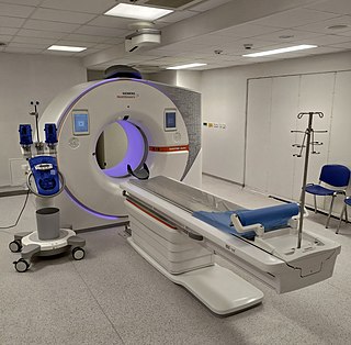
A computed tomography scan is a medical imaging technique used to obtain detailed internal images of the body. The personnel that perform CT scans are called radiographers or radiology technologists.
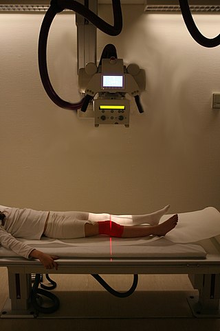
Radiography is an imaging technique using X-rays, gamma rays, or similar ionizing radiation and non-ionizing radiation to view the internal form of an object. Applications of radiography include medical and industrial radiography. Similar techniques are used in airport security,. To create an image in conventional radiography, a beam of X-rays is produced by an X-ray generator and it is projected towards the object. A certain amount of the X-rays or other radiation are absorbed by the object, dependent on the object's density and structural composition. The X-rays that pass through the object are captured behind the object by a detector. The generation of flat two-dimensional images by this technique is called projectional radiography. In computed tomography, an X-ray source and its associated detectors rotate around the subject, which itself moves through the conical X-ray beam produced. Any given point within the subject is crossed from many directions by many different beams at different times. Information regarding the attenuation of these beams is collated and subjected to computation to generate two-dimensional images on three planes which can be further processed to produce a three-dimensional image.
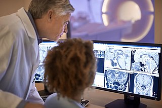
Radiology is the medical specialty that uses medical imaging to diagnose diseases and guide their treatment, within the bodies of humans and other animals. It began with radiography, but today it includes all imaging modalities, including those that use no ionizing electromagnetic radiation, as well as others that do, such as computed tomography (CT), fluoroscopy, and nuclear medicine including positron emission tomography (PET). Interventional radiology is the performance of usually minimally invasive medical procedures with the guidance of imaging technologies such as those mentioned above.

Tomography is imaging by sections or sectioning that uses any kind of penetrating wave. The method is used in radiology, archaeology, biology, atmospheric science, geophysics, oceanography, plasma physics, materials science, cosmochemistry, astrophysics, quantum information, and other areas of science. The word tomography is derived from Ancient Greek τόμος tomos, "slice, section" and γράφω graphō, "to write" or, in this context as well, "to describe." A device used in tomography is called a tomograph, while the image produced is a tomogram.
The Hounsfield scale, named after Sir Godfrey Hounsfield, is a quantitative scale for describing radiodensity. It is frequently used in CT scans, where its value is also termed CT number.
Technicare, formerly known as Ohio Nuclear, made CT, DR and MRI scanners and other medical imaging equipment. Its headquarters was in Solon, Ohio. Originally an independent company which became publicly traded, it was later purchased by Johnson & Johnson. At the time, Invacare was also owned by Technicare. A Harvard Business Case was written about the challenges that precipitated the transition. The company did not do well under Johnson & Johnson and in 1986, under economic pressure following unrelated losses from two Tylenol product tampering cases, J&J folded the company, selling the intellectual property and profitable service business to General Electric, a competitor.
Tomographic reconstruction is a type of multidimensional inverse problem where the challenge is to yield an estimate of a specific system from a finite number of projections. The mathematical basis for tomographic imaging was laid down by Johann Radon. A notable example of applications is the reconstruction of computed tomography (CT) where cross-sectional images of patients are obtained in non-invasive manner. Recent developments have seen the Radon transform and its inverse used for tasks related to realistic object insertion required for testing and evaluating computed tomography use in airport security.
Willi A. Kalender is a German medical physicist and professor and former chairman of the Institute of Medical Physics of the University of Erlangen-Nuremberg. Kalender has produced several new technologies in the field of diagnostic radiology imaging.
Image-guided radiation therapy is the process of frequent imaging, during a course of radiation treatment, used to direct the treatment, position the patient, and compare to the pre-therapy imaging from the treatment plan. Immediately prior to, or during, a treatment fraction, the patient is localized in the treatment room in the same position as planned from the reference imaging dataset. An example of IGRT would include comparison of a cone beam computed tomography (CBCT) dataset, acquired on the treatment machine, with the computed tomography (CT) dataset from planning. IGRT would also include matching planar kilovoltage (kV) radiographs or megavoltage (MV) images with digital reconstructed radiographs (DRRs) from the planning CT.

High-resolution computed tomography (HRCT) is a type of computed tomography (CT) with specific techniques to enhance image resolution. It is used in the diagnosis of various health problems, though most commonly for lung disease, by assessing the lung parenchyma. On the other hand, HRCT of the temporal bone is used to diagnose various middle ear diseases such as otitis media, cholesteatoma, and evaluations after ear operations.

ITK-SNAP is an interactive software application that allows users to navigate three-dimensional medical images, manually delineate anatomical regions of interest, and perform automatic image segmentation. The software was designed with the audience of clinical and basic science researchers in mind, and emphasis has been placed on having a user-friendly interface and maintaining a limited feature set to prevent feature creep. ITK-SNAP is most frequently used to work with magnetic resonance imaging (MRI), cone-beam computed tomography (CBCT) and computed tomography (CT) data sets.

Tomosynthesis, also digital tomosynthesis (DTS), is a method for performing high-resolution limited-angle tomography at radiation dose levels comparable with projectional radiography. It has been studied for a variety of clinical applications, including vascular imaging, dental imaging, orthopedic imaging, mammographic imaging, musculoskeletal imaging, and chest imaging.
Flat-panel Volume CT is a technique under development to make computed tomography images with improved performance. The key difference between volume CT and traditional CT is that volume CT uses a two-dimensional x-ray detector orientation, to take multiple two-dimensional images. On the other hand, the conventional CT uses a one-dimensional x-ray detector orientation to take one-dimensional x-ray images.

Industrial computed tomography (CT) scanning is any computer-aided tomographic process, usually X-ray computed tomography, that uses irradiation to produce three-dimensional internal and external representations of a scanned object. Industrial CT scanning has been used in many areas of industry for internal inspection of components. Some of the key uses for industrial CT scanning have been flaw detection, failure analysis, metrology, assembly analysis and reverse engineering applications. Just as in medical imaging, industrial imaging includes both nontomographic radiography and computed tomographic radiography.

Cone beam computed tomography is a medical imaging technique consisting of X-ray computed tomography where the X-rays are divergent, forming a cone.

X-ray computed tomography operates by using an X-ray generator that rotates around the object; X-ray detectors are positioned on the opposite side of the circle from the X-ray source.
Wojciech (Wojtek) Zbijewski is an American biomedical engineering and medical physics working in the fields of Computed tomography (CT), Cone beam computed tomography (CBCT), image reconstruction in CT, and applications of CT and CBCT in orthopedics. He is faculty at the Department of Biomedical Engineering at Johns Hopkins School of Medicine.
Jeffrey Harold Siewerdsen is an American physicist and biomedical engineer who is a Professor of Imaging Physics at The University of Texas MD Anderson Cancer Center as well as Biomedical Engineering, Computer Science, Radiology, and Neurosurgery at Johns Hopkins University.He is among the original inventors of cone-beam CT-guided radiotherapy as well as weight-bearing cone-beam CT for musculoskeletal radiology and orthopedic surgery. His work also includes the early development of flat-panel detectors on mobile C-arms for intraoperative cone-beam CT in image-guided surgery. He developed early models for the signal and noise performance of flat-panel detectors and later extended such analysis to dual-energy imaging and 3D imaging performance in cone-beam CT. He founded the ISTAR Lab in the Department of Biomedical Engineering, the Carnegie Center for Surgical Innovation at Johns Hopkins Hospital, and the Surgical Data Science Program at the Institute for Data Science in Oncology at The University of Texas MD Anderson Cancer Center.
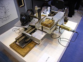
The history of X-ray computed tomography (CT) dates back to at least 1917 with the mathematical theory of the Radon transform. In the early 1900s an Italian radiologist named Alessandro Vallebona invented tomography which used radiographic film to see a single slice of the body. It was not widely used until the 1930s, when Dr Bernard George Ziedses des Plantes developed a practical method for implementing the technique, known as focal plane tomography. It relies on mechanical movement of the X-ray beam source and capture film in unison to ensure that the plane of interest remains in focus with objects falling outside of the plane being examined blurring out.
Ge Wang is a medical imaging scientist focusing on computed tomography (CT) and artificial intelligence (AI) especially deep learning. He is the Clark & Crossan Chair Professor of Biomedical Engineering and the Director of the Biomedical Imaging Center at Rensselaer Polytechnic Institute, Troy, New York, USA. He is known for his pioneering work on CT and AI-based imaging. He is Fellow of American Institute for Medical and Biological Engineering (AIMBE), Institute of Electrical and Electronics Engineers (IEEE), International Society for Optics and Photonics (SPIE), Optical Society of America (OSA/Optica), American Association of Physicists in Medicine (AAPM), American Association for the Advancement of Science (AAAS), and National Academy of Inventors (NAI).
References
- ↑ Defrise, M., Noo, F., Kudo, H. "Physics in Medicine and Biology," 45(3):623-643, 2000.
- ↑ La Riviere, P.J., Crawford, C.R. "Journal of Medical Imaging," 8(5): 052111-1-12, 2021.
- ↑ "Proposed next generation nano-computed tomography system will enhance nanoscale research". Virginia Tech. Retrieved 15 September 2024.
- ↑ Wang, G.; Lin, T.-H.; Cheng, P.; Shinozaki, D.M. (1993). "A general cone-beam reconstruction algorithm". IEEE Transactions on Medical Imaging. 12 (3): 486–496. doi:10.1109/42.241876. PMID 18218441.
- ↑ A. Katsevich. Theoretically exact filtered backprojection-type inversion algorithm for Spiral CT. SIAM J. Appl. Math., 62 (2002), pp. 2012-2026
- ↑ Anderson, Julia (13 October 2016). "- A Prize From a King". College of Sciences News.
- ↑ "Medical Imaging in Increasing Dimensions". American Scientist. 10 August 2023.
- ↑ Lv Y, Katsevich A, Zhao J, Yu H, Wang G: Fast exact/quasi-exact FBP algorithms for triple-source helical cone-beam CT. IEEE Trans. Medical Imaging 29:756-770, 2010
- ↑ Verhoeven, Roel L. J.; Kops, Stephan E. P.; Wijma, Inge N.; ter Woerds, Desi K. M.; van der Heijden, Erik H. F. M. (30 August 2023). "Cone-beam CT in lung biopsy: a clinical practice review on lessons learned and future perspectives". Annals of Translational Medicine. 11 (10): 361. doi: 10.21037/atm-22-2845 . ISSN 2305-5839. PMC 10477635 . PMID 37675336.
- ↑ Bapst, Blanche; Lagadec, Matthieu; Breguet, Romain; Vilgrain, Valérie; Ronot, Maxime (January 2016). "Cone Beam Computed Tomography (CBCT) in the Field of Interventional Oncology of the Liver". CardioVascular and Interventional Radiology. 39 (1): 8–20. doi:10.1007/s00270-015-1180-6. ISSN 1432-086X. PMID 26178776.
- ↑ Hu, H., Duerinckx, A. J., Foley, W. D., & Cooper, C. (2000). Helical/spiral CT in cardiovascular disease. Journal of Thoracic Imaging, 15(4), 290-305. doi:10.1097/00005382-200010000-00004
- ↑ Hutchison, Chad. "5 Advantages of Using CBCT (Cone Beam CT) in Orthopedics". mavenimaging.com. Retrieved 14 September 2024.
- ↑ Wang, X., Wu, Z., & Liu, Y. (2019). The clinical application of cone-beam computed tomography in emergency trauma. Journal of Clinical Medicine Research, 11(7), 484-491. doi:10.14740/jocmr3844
- ↑ Key, Brandon M.; Tutton, Sean M.; Scheidt, Matthew J. (July 2023). "Cone-Beam CT With Enhanced Needle Guidance and Augmented Fluoroscopy Overlay: Applications in Interventional Radiology". American Journal of Roentgenology. 221 (1): 92–101. doi:10.2214/AJR.22.28712. ISSN 0361-803X. PMID 37095661.