Related Research Articles

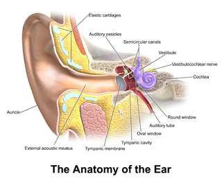
The middle ear is the portion of the ear medial to the eardrum, and distal to the oval window of the cochlea.
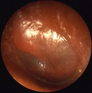
In the anatomy of humans and various other tetrapods, the eardrum, also called the tympanic membrane or myringa, is a thin, cone-shaped membrane that separates the external ear from the middle ear. Its function is to transmit sound from the air to the ossicles inside the middle ear, and then to the oval window in the fluid-filled cochlea. Hence, it ultimately converts and amplifies vibration in the air to vibration in cochlear fluid. The malleus bone bridges the gap between the eardrum and the other ossicles.
Otitis media is a group of inflammatory diseases of the middle ear. One of the two main types is acute otitis media (AOM), an infection of rapid onset that usually presents with ear pain. In young children this may result in pulling at the ear, increased crying, and poor sleep. Decreased eating and a fever may also be present. The other main type is otitis media with effusion (OME), typically not associated with symptoms, although occasionally a feeling of fullness is described; it is defined as the presence of non-infectious fluid in the middle ear which may persist for weeks or months often after an episode of acute otitis media. Chronic suppurative otitis media (CSOM) is middle ear inflammation that results in a perforated tympanic membrane with discharge from the ear for more than six weeks. It may be a complication of acute otitis media. Pain is rarely present. All three types of otitis media may be associated with hearing loss. If children with hearing loss due to OME do not learn sign language, it may affect their ability to learn.

In anatomy, the Eustachian tube, also known as the auditory tube or pharyngotympanic tube, is a tube that links the nasopharynx to the middle ear, of which it is also a part. In adult humans, the Eustachian tube is approximately 35 mm (1.4 in) long and 3 mm (0.12 in) in diameter. It is named after the sixteenth-century Italian anatomist Bartolomeo Eustachi.
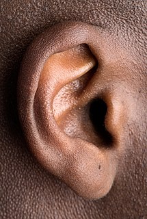
The ear is the organ that enables hearing and, in mammals, body balance using the vestibular system. In mammals, the ear is usually described as having three parts—the outer ear, the middle ear and the inner ear. The outer ear consists of the pinna and the ear canal. Since the outer ear is the only visible portion of the ear in most animals, the word "ear" often refers to the external part alone. The middle ear includes the tympanic cavity and the three ossicles. The inner ear sits in the bony labyrinth, and contains structures which are key to several senses: the semicircular canals, which enable balance and eye tracking when moving; the utricle and saccule, which enable balance when stationary; and the cochlea, which enables hearing. The ears of vertebrates are placed somewhat symmetrically on either side of the head, an arrangement that aids sound localisation.

Conductive hearing loss (CHL) occurs when there is a problem transferring sound waves anywhere along the pathway through the outer ear, tympanic membrane (eardrum), or middle ear (ossicles). If a conductive hearing loss occurs in conjunction with a sensorineural hearing loss, it is referred to as a mixed hearing loss. Depending upon the severity and nature of the conductive loss, this type of hearing impairment can often be treated with surgical intervention or pharmaceuticals to partially or, in some cases, fully restore hearing acuity to within normal range. However, cases of permanent or chronic conductive hearing loss may require other treatment modalities such as hearing aid devices to improve detection of sound and speech perception.
A myringotomy is a surgical procedure in which an incision is created in the eardrum to relieve pressure caused by excessive buildup of fluid, or to drain pus from the middle ear. A tympanostomy tube may be inserted through the eardrum to keep the middle ear aerated for a prolonged time and to prevent reaccumulation of fluid. Without the insertion of a tube, the incision usually heals spontaneously within two to three weeks. Depending on the type, the tube is either naturally extruded in 6 to 12 months or removed during a minor procedure.

Tympanoplasty is the surgical operation performed to reconstruct hearing mechanism of middle ear

Ear pain, also known as earache or otalgia, is pain in the ear. Primary ear pain is pain that originates from the ear. Secondary ear pain is a type of referred pain, meaning that the source of the pain differs from the location where the pain is felt.
Patulous Eustachian tube (PET) is the name of a physical disorder where the Eustachian tube, which is normally closed, instead stays intermittently open. When this occurs, the person experiences autophony, the hearing of self-generated sounds. These sounds, such as one's own breathing, voice, and heartbeat, vibrate directly onto the ear drum and can create a "bucket on the head" effect. PET is a form of eustachian tube dysfunction (ETD), which is said to be present in about 1 percent of the general population.

Tympanostomy tube, also known as a grommet or myringotomy tube, is a small tube inserted into the eardrum in order to keep the middle ear aerated for a prolonged period of time, and to prevent the accumulation of fluid in the middle ear. The operation to insert the tube involves a myringotomy and is performed under local or general anesthesia. The tube itself is made in a variety of designs. The most commonly used type is shaped like a grommet. When it is necessary to keep the middle ear ventilated for a very long period, a "T"-shaped tube may be used, as these "T-tubes" can stay in place for 2–4 years. Materials used to construct the tube are most often plastics such as silicone or Teflon. Stainless steel tubes exist, but are no longer in frequent use.
Tympanocentesis is the drainage of fluid from the middle ear usually caused by otitis media, by using a small-gauge needle to puncture the tympanic membrane, also known as the eardrum.

Tympanometry is an acoustic evaluation of the condition of the middle ear eardrum and the conduction bones by creating variations of air pressure in the ear canal.

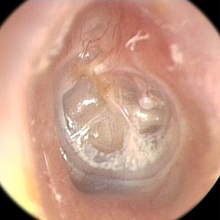
An otoscope or auriscope is a medical device which is used to look into the ears. Health care providers use otoscopes to screen for illness during regular check-ups and also to investigate ear symptoms. An otoscope potentially gives a view of the ear canal and tympanic membrane or eardrum. Because the eardrum is the border separating the external ear canal from the middle ear, its characteristics can be indicative of various diseases of the middle ear space. The presence of earwax (cerumen), shed skin, pus, canal skin edema, foreign body, and various ear diseases can obscure any view of the eardrum and thus severely compromise the value of otoscopy done with a common otoscope, but confirm the presence of obstructing symptoms.

A perforated eardrum is a hole in the eardrum. It can be caused by infection, trauma, overpressure, inappropriate ear clearing, and changes in middle ear pressure. An otoscope can be used to view the eardrum to diagnose a perforation. Perforations may heal naturally, or require surgery.
Otomycosis is a fungal ear infection, a superficial mycotic infection of the outer ear canal. It is more common in tropical countries. The infection may be either subacute or acute and is characterized by malodorous discharge, inflammation, pruritus, scaling, and severe discomfort. The mycosis results in inflammation, superficial epithelial exfoliation, masses of debris containing hyphae, suppuration, and pain.

Tympanosclerosis is a condition caused by hyalinization and subsequent calcification of subepithelial connective tissue of the tympanic membrane and middle ear, sometimes resulting in a detrimental effect to hearing.
Tympanic membrane retraction describes a condition in which a part of the eardrum lies deeper within the ear than its normal position.
Granular myringitis is a long term condition in which there is inflammation of the tympanic membrane in the ear and formation of granulation tissue within the tympanic membrane. It is a type of otitis externa.

The malleus, or hammer, is a hammer-shaped small bone or ossicle of the middle ear. It connects with the incus, and is attached to the inner surface of the eardrum. The word is Latin for 'hammer' or 'mallet'. It transmits the sound vibrations from the eardrum to the incus (anvil).
References
- ↑ Stanley Gelfand (19 March 2009). Essentials of Audiology. Thieme. p. 40. ISBN 978-1-60406-155-0.
- ↑ Cone of Light Anatomy - "..:: The Tympanic Membrane ::". Archived from the original on 2012-04-05. Retrieved 2011-12-24.
- ↑ Advanced Therapy in Otitis Media. PMPH-USA. 2004. p. 56. ISBN 978-1-55009-201-1.
- ↑ Robert Thayer Sataloff; Joseph Sataloff (24 April 2006). Occupational Hearing Loss, Third Edition. CRC Press. p. 38. ISBN 978-1-4200-1547-8.