DNA laddering

Last updated
DNA laddering (left) visualised in an agarose gel by ethidium bromide staining. A 1 kb marker (middle) and control DNA (right) are included. Apoptotic DNA Laddering.png
DNA laddering (left) visualised in an agarose gel by ethidium bromide staining. A 1 kb marker (middle) and control DNA (right) are included.

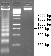
DNA laddering is a feature that can be observed when DNA fragments, resulting from Apoptosis DNA fragmentation are visualized after separation by gel electrophoresis the first described in 1980 by Andrew Wyllie at the University Edinburgh medical school [1] DNA fragments can also be delected in cells that underwent necrosis, when theses DNA fragments after separation are subjected to gel electrophoresis which in the results in a characteristic ladder pattern,

Contents

DNA degradation

DNA laddering is a distinctive feature of DNA degraded by caspase-activated DNase (CAD), which is a key event during apoptosis. CAD cleaves genomic DNA at internucleosomal linker regions, resulting in DNA fragments that are multiples of 180–185 base-pairs in length. [2] Separation of the fragments by agarose gel electrophoresis and subsequent visualization, for example by ethidium bromide staining, results in a characteristic "ladder" pattern. A simple method of selective extraction of fragmented DNA from apoptotic cells without the presence of high molecular weight DNA sections, generating the laddering pattern, utilizes pretreatment of cells in ethanol. [3]

Apoptosis and necrosis

While most of the morphological features of apoptotic cells are short-lived, DNA laddering can be used as final state read-out method and has therefore become a reliable method to distinguish apoptosis from necrosis. [4] DNA laddering can also be used to see if cells underwent apoptosis in the presence of a virus. [5] This is useful because it can help determine the effects a virus has on a cell.

DNA laddering can only be used to detect apoptosis during the later stages of apoptosis. This is due to DNA fragmentation taking place in a later stage of the apoptosis process. [2] DNA laddering is used to test for apoptosis of many cells, and is not accurate at testing for only a few cells that committed apoptosis. [2] To enhance the accuracy in testing for apoptosis, other assays are used along with DNA laddering such as TEM and TUNEL. [2] With recent improvements to DNA laddering, DNA laddering has become a more reliable, and reasonable technique to use when detecting apoptosis. [6] It is also important to note that DNA laddering occurs differently depending on the type of cell, so there may be slight changes in the process of DNA laddering depending on the cell that is being investigated. [7]

See also

Related Research Articles

<span class="mw-page-title-main">Apoptosis</span> Programmed cell death in multicellular organisms

Apoptosis is a form of programmed cell death that occurs in multicellular organisms and in some eukaryotic, single-celled microorganisms such as yeast. Biochemical events lead to characteristic cell changes (morphology) and death. These changes include blebbing, cell shrinkage, nuclear fragmentation, chromatin condensation, DNA fragmentation, and mRNA decay. The average adult human loses between 50 and 70 billion cells each day due to apoptosis. For an average human child between eight and fourteen years old, each day the approximate loss is 20 to 30 billion cells.

<span class="mw-page-title-main">Gel electrophoresis</span> Method for separation and analysis of biomolecules

Gel electrophoresis is a method for separation and analysis of biomacromolecules and their fragments, based on their size and charge. It is used in clinical chemistry to separate proteins by charge or size and in biochemistry and molecular biology to separate a mixed population of DNA and RNA fragments by length, to estimate the size of DNA and RNA fragments or to separate proteins by charge.

Programmed cell death is the death of a cell as a result of events inside of a cell, such as apoptosis or autophagy. PCD is carried out in a biological process, which usually confers advantage during an organism's lifecycle. For example, the differentiation of fingers and toes in a developing human embryo occurs because cells between the fingers apoptose; the result is that the digits are separate. PCD serves fundamental functions during both plant and animal tissue development.

Deoxyribonuclease refers to a group of glycoprotein endonucleases which are enzymes that catalyze the hydrolytic cleavage of phosphodiester linkages in the DNA backbone, thus degrading DNA. The role of the DNase enzyme in cells includes breaking down extracellular DNA (ecDNA) excreted by apoptosis, necrosis, and neutrophil extracellular traps (NET) of cells to help reduce inflammatory responses that otherwise are elicited. A wide variety of deoxyribonucleases are known and fall into one of two families, which differ in their substrate specificities, chemical mechanisms, and biological functions. Laboratory applications of DNase include purifying proteins when extracted from prokaryotic organisms. Additionally, DNase has been applied as a treatment for diseases that are caused by ecDNA in the blood plasma. Assays of DNase are emerging in the research field as well.

A DNase footprinting assay is a DNA footprinting technique from molecular biology/biochemistry that detects DNA-protein interaction using the fact that a protein bound to DNA will often protect that DNA from enzymatic cleavage. This makes it possible to locate a protein binding site on a particular DNA molecule. The method uses an enzyme, deoxyribonuclease, to cut the radioactively end-labeled DNA, followed by gel electrophoresis to detect the resulting cleavage pattern.

In the field of genetics, a suicide gene is a gene that will cause a cell to kill itself through the process of apoptosis. Activation of a suicide gene can cause death through a variety of pathways, but one important cellular "switch" to induce apoptosis is the p53 protein. Stimulation or introduction of suicide genes is a potential way of treating cancer or other proliferative diseases.

DNA fragmentation is the separation or breaking of DNA strands into pieces. It can be done intentionally by laboratory personnel or by cells, or can occur spontaneously. Spontaneous or accidental DNA fragmentation is fragmentation that gradually accumulates in a cell. It can be measured by e.g. the Comet assay or by the TUNEL assay.

<span class="mw-page-title-main">TUNEL assay</span>

Terminal deoxynucleotidyl transferase dUTP nick end labeling (TUNEL) is a method for detecting DNA fragmentation by labeling the 3′- hydroxyl termini in the double-strand DNA breaks generated during apoptosis.

<span class="mw-page-title-main">Electrophoretic mobility shift assay</span>

An electrophoretic mobility shift assay (EMSA) or mobility shift electrophoresis, also referred as a gel shift assay, gel mobility shift assay, band shift assay, or gel retardation assay, is a common affinity electrophoresis technique used to study protein–DNA or protein–RNA interactions. This procedure can determine if a protein or mixture of proteins is capable of binding to a given DNA or RNA sequence, and can sometimes indicate if more than one protein molecule is involved in the binding complex. Gel shift assays are often performed in vitro concurrently with DNase footprinting, primer extension, and promoter-probe experiments when studying transcription initiation, DNA gang replication, DNA repair or RNA processing and maturation, as well as pre-mRNA splicing. Although precursors can be found in earlier literature, most current assays are based on methods described by Garner and Revzin and Fried and Crothers.

DNA footprinting is a method of investigating the sequence specificity of DNA-binding proteins in vitro. This technique can be used to study protein-DNA interactions both outside and within cells.

<span class="mw-page-title-main">Comet assay</span>

The single cell gel electrophoresis assay is an uncomplicated and sensitive technique for the detection of DNA damage at the level of the individual eukaryotic cell. It was first developed by Östling & Johansson in 1984 and later modified by Singh et al. in 1988. It has since increased in popularity as a standard technique for evaluation of DNA damage/repair, biomonitoring and genotoxicity testing. It involves the encapsulation of cells in a low-melting-point agarose suspension, lysis of the cells in neutral or alkaline (pH>13) conditions, and electrophoresis of the suspended lysed cells. The term "comet" refers to the pattern of DNA migration through the electrophoresis gel, which often resembles a comet.

Fragmentation describes the process of splitting into several pieces or fragments. In cell biology, fragmentation is useful for a cell during both DNA cloning and apoptosis. DNA cloning is important in asexual reproduction or creation of identical DNA molecules, and can be performed spontaneously by the cell or intentionally by laboratory researchers. Apoptosis is the programmed destruction of cells, and the DNA molecules within them, and is a highly regulated process. These two ways in which fragmentation is used in cellular processes describe normal cellular functions and common laboratory procedures performed with cells. However, problems within a cell can sometimes cause fragmentation that results in irregularities such as red blood cell fragmentation and sperm cell DNA fragmentation.

<span class="mw-page-title-main">DFFA</span> Protein-coding gene in the species Homo sapiens

DNA fragmentation factor subunit alpha (DFFA), also known as Inhibitor of caspase-activated DNase (ICAD), is a protein that in humans is encoded by the DFFA gene.

<span class="mw-page-title-main">ENDOG</span> Protein-coding gene in the species Homo sapiens

Endonuclease G, mitochondrial is an enzyme that in humans is encoded by the ENDOG gene. This protein primarily participates in caspase-independent apoptosis via DNA degradation when translocating from the mitochondrion to nucleus under oxidative stress. As a result, EndoG has been implicated in cancer, aging, and neurodegenerative diseases such as Parkinson’s disease (PD). Regulation of its expression levels thus holds potential to treat or ameliorate those conditions.

<span class="mw-page-title-main">Apoptotic DNA fragmentation</span> Cleavage of DNA into tiny pieces during apoptosis

Apoptotic DNA fragmentation is a key feature of apoptosis, a type of programmed cell death. Apoptosis is characterized by the activation of endogenous endonucleases, particularly the caspase-3 activated DNase (CAD), with subsequent cleavage of nuclear DNA into internucleosomal fragments of roughly 180 base pairs (bp) and multiples thereof (360, 540 etc.). The apoptotic DNA fragmentation is being used as a marker of apoptosis and for identification of apoptotic cells either via the DNA laddering assay, the TUNEL assay, or the by detection of cells with fractional DNA content ("sub G1 cells") on DNA content frequency histograms e.g. as in the Nicoletti assay.

M30 Apoptosense® ELISA is an enzyme-linked immunosorbent assay developed for the detection of soluble caspase-cleaved keratin 18.

<span class="mw-page-title-main">Caspase-activated DNase</span> Protein-coding gene in the species Homo sapiens

Caspase-activated DNase (CAD) or DNA fragmentation factor subunit beta is a protein that in humans is encoded by the DFFB gene. It breaks up the DNA during apoptosis and promotes cell differentiation. It is usually an inactive monomer inhibited by ICAD. This is cleaved before dimerization.

Ischemic cell death, or oncosis, is a form of accidental cell death. The process is characterized by an ATP depletion within the cell leading to impairment of ionic pumps, cell swelling, clearing of the cytosol, dilation of the endoplasmic reticulum and golgi apparatus, mitochondrial condensation, chromatin clumping, and cytoplasmic bleb formation. Oncosis refers to a series of cellular reactions following injury that precedes cell death. The process of oncosis is divided into three stages. First, the cell becomes committed to oncosis as a result of damage incurred to the plasma membrane through toxicity or ischemia, resulting in the leak of ions and water due to ATP depletion. The ionic imbalance that occurs subsequently causes the cell to swell without a concurrent change in membrane permeability to reverse the swelling. In stage two, the reversibility threshold for the cell is passed and the cell becomes committed to cell death. During this stage the membrane becomes abnormally permeable to trypan blue and propidium iodide, indicating membrane compromise. The final stage is cell death and removal of the cell via phagocytosis mediated by an inflammatory response.

<span class="mw-page-title-main">Necroptosis</span> Programmed form of necrosis, or inflammatory cell death

Necroptosis is a programmed form of necrosis, or inflammatory cell death. Conventionally, necrosis is associated with unprogrammed cell death resulting from cellular damage or infiltration by pathogens, in contrast to orderly, programmed cell death via apoptosis. The discovery of necroptosis showed that cells can execute necrosis in a programmed fashion and that apoptosis is not always the preferred form of cell death. Furthermore, the immunogenic nature of necroptosis favors its participation in certain circumstances, such as aiding in defence against pathogens by the immune system. Necroptosis is well defined as a viral defense mechanism, allowing the cell to undergo "cellular suicide" in a caspase-independent fashion in the presence of viral caspase inhibitors to restrict virus replication. In addition to being a response to disease, necroptosis has also been characterized as a component of inflammatory diseases such as Crohn's disease, pancreatitis, and myocardial infarction.

Urinary cell-free DNA (ucfDNA) refers to DNA fragments in urine released by urogenital and non-urogenital cells. Shed cells on urogenital tract release high- or low-molecular-weight DNA fragments via apoptosis and necrosis, while circulating cell-free DNA (cfDNA) that passes through glomerular pores contributes to low-molecular-weight DNA. Most of the ucfDNA is low-molecular-weight DNA in the size of 150-250 base pairs. The detection of ucfDNA composition allows the quantification of cfDNA, circulating tumour DNA, and cell-free fetal DNA components. Many commercial kits and devices have been developed for ucfDNA isolation, quantification, and quality assessment.

References

  1. Kressel, M.; Groscurth, P. (1994-12-01). "Distinction of apoptotic and necrotic cell death by in situ labelling of fragmented DNA". Cell and Tissue Research. 278 (3): 549–556. doi:10.1007/s004410050244. ISSN   0302-766X. PMID   7850865.
  2. 1 2 3 4 Elmore, Susan (2007-01-01). "Apoptosis: A Review of Programmed Cell Death". Toxicologic Pathology. 35 (4): 495–516. doi:10.1080/01926230701320337. ISSN   0192-6233. PMC   2117903 . PMID   17562483.
  3. Gong, J.; Traganos, F.; Darzynkiewicz, Z. (1994). "A selective procedure for DNA extraction from apoptotic cells applicable for gel electrophoresis and flow cytometry". Analytical Biochemistry. 218 (2): 314–9. doi:10.1006/abio.1994.1184. PMID   8074286.
  4. Iwata M, Myerson D, Torok-Storb B, Zager RA (December 1994). "An evaluation of renal tubular DNA laddering in response to oxygen deprivation and oxidant injury". Journal of the American Society of Nephrology . 5 (6): 1307–1313. doi: 10.1681/ASN.V561307 . ISSN   1046-6673. PMID   7893995.
  5. Srivastava, Vikram; Rawall, Shweta; Vijayan, V. K.; Khanna, Madhu (2009-05-01). "Influenza a virus induced apoptosis: inhibition of DNA laddering & caspase-3 activity by zinc supplementation in cultured HeLa cells". The Indian Journal of Medical Research. 129 (5): 579–586. ISSN   0971-5916. PMID   19675388.
  6. Rahbar Saadat, Yalda; Saeidi, Nazli; Zununi Vahed, Sepideh; Barzegari, Abolfazl; Barar, Jaleh (2015-01-01). "An update to DNA ladder assay for apoptosis detection". BioImpacts. 5 (1): 25–28. doi:10.15171/bi.2015.01. ISSN   2228-5652. PMC   4401164 . PMID   25901294.
  7. Jiang, Ai-Liang; Cheng, Yanwei; Li, Jianyou; Zhang, Wei (2008-07-31). "A zinc-dependent nuclear endonuclease is responsible for DNA laddering during salt-induced programmed cell death in root tip cells of rice". Journal of Plant Physiology. 165 (11): 1134–1141. doi:10.1016/j.jplph.2007.12.008. ISSN   1618-1328. PMID   18295371.