Related Research Articles

An electron microscope is a microscope that uses a beam of electrons as a source of illumination. They use electron optics that are analogous to the glass lenses of an optical light microscope to control the electron beam, for instance focusing them to produce magnified images or electron diffraction patterns. As the wavelength of an electron can be up to 100,000 times smaller than that of visible light, electron microscopes have a much higher resolution of about 0.1 nm, which compares to about 200 nm for light microscopes. Electron microscope may refer to:

Histology, also known as microscopic anatomy or microanatomy, is the branch of biology that studies the microscopic anatomy of biological tissues. Histology is the microscopic counterpart to gross anatomy, which looks at larger structures visible without a microscope. Although one may divide microscopic anatomy into organology, the study of organs, histology, the study of tissues, and cytology, the study of cells, modern usage places all of these topics under the field of histology. In medicine, histopathology is the branch of histology that includes the microscopic identification and study of diseased tissue. In the field of paleontology, the term paleohistology refers to the histology of fossil organisms.

Pathology is the study of disease and injury. The word pathology also refers to the study of disease in general, incorporating a wide range of biology research fields and medical practices. However, when used in the context of modern medical treatment, the term is often used in a narrower fashion to refer to processes and tests that fall within the contemporary medical field of "general pathology", an area that includes a number of distinct but inter-related medical specialties that diagnose disease, mostly through analysis of tissue and human cell samples. Idiomatically, "a pathology" may also refer to the predicted or actual progression of particular diseases, and the affix pathy is sometimes used to indicate a state of disease in cases of both physical ailment and psychological conditions. A physician practicing pathology is called a pathologist.

Transmission electron microscopy (TEM) is a microscopy technique in which a beam of electrons is transmitted through a specimen to form an image. The specimen is most often an ultrathin section less than 100 nm thick or a suspension on a grid. An image is formed from the interaction of the electrons with the sample as the beam is transmitted through the specimen. The image is then magnified and focused onto an imaging device, such as a fluorescent screen, a layer of photographic film, or a sensor such as a scintillator attached to a charge-coupled device.

Staining is a technique used to enhance contrast in samples, generally at the microscopic level. Stains and dyes are frequently used in histology, in cytology, and in the medical fields of histopathology, hematology, and cytopathology that focus on the study and diagnoses of diseases at the microscopic level. Stains may be used to define biological tissues, cell populations, or organelles within individual cells.

In biochemistry, immunostaining is any use of an antibody-based method to detect a specific protein in a sample. The term "immunostaining" was originally used to refer to the immunohistochemical staining of tissue sections, as first described by Albert Coons in 1941. However, immunostaining now encompasses a broad range of techniques used in histology, cell biology, and molecular biology that use antibody-based staining methods.
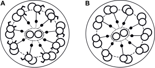
Primary ciliary dyskinesia (PCD) is a rare, autosomal recessive genetic ciliopathy, that causes defects in the action of cilia lining the upper and lower respiratory tract, sinuses, Eustachian tube, middle ear, Fallopian tube, and flagella of sperm cells. The alternative name of "immotile ciliary syndrome" is no longer favored as the cilia do have movement, but are merely inefficient or unsynchronized. When accompanied by situs inversus the condition is known as Kartagener syndrome.

Histopathology refers to the microscopic examination of tissue in order to study the manifestations of disease. Specifically, in clinical medicine, histopathology refers to the examination of a biopsy or surgical specimen by a pathologist, after the specimen has been processed and histological sections have been placed onto glass slides. In contrast, cytopathology examines free cells or tissue micro-fragments.
A microtome is a cutting tool used to produce extremely thin slices of material known as sections, with the process being termed microsectioning. Important in science, microtomes are used in microscopy for the preparation of samples for observation under transmitted light or electron radiation.
Ultramicrotomy is a method for cutting specimens into extremely thin slices, called ultra-thin sections, that can be studied and documented at different magnifications in a transmission electron microscope (TEM). It is used mostly for biological specimens, but sections of plastics and soft metals can also be prepared. Sections must be very thin because the 50 to 125 kV electrons of the standard electron microscope cannot pass through biological material much thicker than 150 nm. For best resolutions, sections should be from 30 to 60 nm. This is roughly the equivalent to splitting a 0.1 mm-thick human hair into 2,000 slices along its diameter, or cutting a single red blood cell into 100 slices.
The frozen section procedure is a pathological laboratory procedure to perform rapid microscopic analysis of a specimen. It is used most often in oncological surgery. The technical name for this procedure is cryosection. The microtome device that cold cuts thin blocks of frozen tissue is called a cryotome.

Uranyl acetate is the acetate salt of uranium oxide, a toxic yellow-green powder useful in certain laboratory tests. Structurally, it is a coordination polymer with formula UO2(CH3CO2)2(H2O)·H2O.
In microscopy, negative staining is an established method, often used in diagnostic microscopy, for contrasting a thin specimen with an optically opaque fluid. In this technique, the background is stained, leaving the actual specimen untouched, and thus visible. This contrasts with positive staining, in which the actual specimen is stained.

Immunolabeling is a biochemical process that enables the detection and localization of an antigen to a particular site within a cell, tissue, or organ. Antigens are organic molecules, usually proteins, capable of binding to an antibody. These antigens can be visualized using a combination of antigen-specific antibody as well as a means of detection, called a tag, that is covalently linked to the antibody. If the immunolabeling process is meant to reveal information about a cell or its substructures, the process is called immunocytochemistry. Immunolabeling of larger structures is called immunohistochemistry.

A diamond knife is a very sharp knife in which the edge is made from diamond, invented by Humberto Fernández-Morán in 1955. Diamond knives are used for medical and scientific applications where an extremely sharp and long-lasting edge is essential. The knives are very expensive to purchase, depending on the quality and size of the knife; in addition the knives must be professionally sharpened as the edge dulls.
Low-voltage electron microscope (LVEM) is an electron microscope which operates at accelerating voltages of a few kiloelectronvolts or less. Traditional electron microscopes use accelerating voltages in the range of 10-1000 keV.

Immunogold labeling or Immunogold staining (IGS) is a staining technique used in electron microscopy. This staining technique is an equivalent of the indirect immunofluorescence technique for visible light. Colloidal gold particles are most often attached to secondary antibodies which are in turn attached to primary antibodies designed to bind a specific antigen or other cell component. Gold is used for its high electron density which increases electron scatter to give high contrast 'dark spots'.
Serial block-face scanning electron microscopy is a method to generate high resolution three-dimensional images from small samples. The technique was developed for brain tissue, but it is widely applicable for any biological samples. A serial block-face scanning electron microscope consists of an ultramicrotome mounted inside the vacuum chamber of a scanning electron microscope. Samples are prepared by methods similar to that in transmission electron microscopy (TEM), typically by fixing the sample with aldehyde, staining with heavy metals such as osmium and uranium then embedding in an epoxy resin. The surface of the block of resin-embedded sample is imaged by detection of back-scattered electrons. Following imaging the ultramicrotome is used to cut a thin section from the face of the block. After the section is cut, the sample block is raised back to the focal plane and imaged again. This sequence of sample imaging, section cutting and block raising can acquire many thousands of images in perfect alignment in an automated fashion. Practical serial block-face scanning electron microscopy was invented in 2004 by Winfried Denk at the Max-Planck-Institute in Heidelberg and is commercially available from Gatan Inc., Thermo Fisher Scientific (VolumeScope) and ConnectomX.
Durcupan is a water-soluble epoxy resin produced by the Fluka subsidiary of Sigma-Aldrich. It is commonly used for embedding electron microscope samples in plastic so they may be sectioned with a microtome and then imaged.

Immune electron microscopy is the equivalent of immunofluorescence, but it uses electron microscopy rather than light microscopy. Immunoelectron microscopy identifies and localizes a molecule of interest, specifically a protein of interest, by attaching it to a particular antibody. This bond can form before or after embedding the cells into slides. A reaction occurs between the antigen and antibody, causing this label to become visible under the microscope. Scanning electron microscopy is a viable option if the antigen is on the surface of the cell, but transmission electron microscopy may be needed to see the label if the antigen is within the cell.
References
- ↑ Shoemark, Amelia; Boon, Mieke; Brochhausen, Christoph; Bukowy-Bieryllo, Zuzanna; Santi, Maria M. De; Goggin, Patricia; Griffin, Paul; Hegele, Richard G.; Hirst, Robert A.; Leigh, Margaret W.; Lupton, Alison; MacKenney, Karen; Omran, Heymut; Pache, Jean-Claude; Pinto, Andreia (2020-04-01). "International consensus guideline for reporting transmission electron microscopy results in the diagnosis of primary ciliary dyskinesia (BEAT PCD TEM Criteria)". European Respiratory Journal. 55 (4). doi: 10.1183/13993003.00725-2019 . ISSN 0903-1936. PMID 32060067.
- ↑ Woods AE, Stirling JW. 2008. Electron microscopy. In, Theory and Practice of Histological Techniques. Eds, Bancroft JD and Gamble M. 6th edition. Churchill Livingstone: pages 601-640
- ↑ Stirling, John; Curry, Alan; Eyden, Brian (2013). Diagnostic Electron Microscopy. A Practical Guide to Interpretation and Technique (1 ed.). Chichester, UK: John Wiley & Sons Ltd. ISBN 978-1-119-97399-7.