
The peripheral nervous system (PNS) is one of two components that make up the nervous system of bilateral animals, with the other part being the central nervous system (CNS). The PNS consists of the nerves and ganglia outside the brain and spinal cord. The main function of the PNS is to connect the CNS to the limbs and organs, essentially serving as a relay between the brain and spinal cord and the rest of the body. Unlike the CNS, the PNS is not protected by the vertebral column and skull, or by the blood–brain barrier, which leaves it exposed to toxins and mechanical injuries.

The autonomic nervous system (ANS), formerly the vegetative nervous system, is a division of the peripheral nervous system that supplies smooth muscle and glands, and thus influences the function of internal organs. The autonomic nervous system is a control system that acts largely unconsciously and regulates bodily functions such as the heart rate, digestion, respiratory rate, pupillary response, urination, and sexual arousal. This system is the primary mechanism in control of the fight-or-flight response.
The parasympathetic nervous system (PSNS) is one of the two divisions of the autonomic nervous system, the other being the sympathetic nervous system. The autonomic nervous system is responsible for regulating the body's unconscious actions. The parasympathetic system is responsible for stimulation of "rest-and-digest" or "feed and breed" activities that occur when the body is at rest, especially after eating, including sexual arousal, salivation, lacrimation (tears), urination, digestion and defecation. Its action is described as being complementary to that of the sympathetic nervous system, which is responsible for stimulating activities associated with the fight-or-flight response.
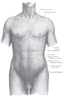
The torso or trunk is an anatomical term for the central part or core of many animal bodies from which extend the neck and limbs. The torso includes: the thoracic segment of the trunk, the abdominal segment of the trunk, and the perineum.
Electroneuronography or electroneurography (ENoG) is a neurological non-invasive test used to study the facial nerve in cases of muscle weakness in one side of the face. The technique of electroneuronography was first used by Esslen and Fisch in 1979 to describe a technique that examines the integrity and conductivity of peripheral nerves. In modern use, ENoG is used to describe study of the facial nerve, while the term nerve conduction study is employed for other nerves.
Neurotmesis is part of Seddon's classification scheme used to classify nerve damage. It is the most serious nerve injury in the scheme. In this type of injury, both the nerve and the nerve sheath are disrupted. While partial recovery may occur, complete recovery is impossible.
Axonotmesis is an injury to the peripheral nerve of one of the extremities of the body. The axons and their myelin sheath are damaged in this kind of injury, but the endoneurium, perineurium and epineurium remain intact. Motor and sensory functions distal to the point of injury are completely lost over time leading to Wallerian Degeneration due to ischemia, or loss of blood supply. Axonotmesis is usually the result of a more severe crush or contusion than neurapraxia.
Neurapraxia is a disorder of the peripheral nervous system in which there is a temporary loss of motor and sensory function due to blockage of nerve conduction, usually lasting an average of six to eight weeks before full recovery. Neurapraxia is derived from the word apraxia, meaning “loss or impairment of the ability to execute complex coordinated movements without muscular or sensory impairment”.

A brachial plexus injury (BPI), also known as brachial plexus lesion, is an injury to the brachial plexus, the network of nerves that conducts signals from the spinal cord to the shoulder, arm and hand. These nerves originate in the fifth, sixth, seventh and eighth cervical (C5–C8), and first thoracic (T1) spinal nerves, and innervate the muscles and skin of the chest, shoulder, arm and hand.
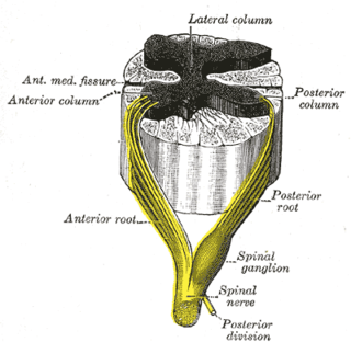
The lateral grey column is one of the three grey columns of the spinal cord ; the others being the anterior and posterior grey columns. The lateral grey column is primarily involved with activity in the sympathetic division of the autonomic motor system. It projects to the side as a triangular field in the thoracic and upper lumbar regions of the postero-lateral part of the anterior grey column.

Augustus Volney Waller FRS was a British neurophysiologist. He was the first to describe the degeneration of severed nerve fibers, now known as Wallerian degeneration.

Nerve injury is injury to nervous tissue. There is no single classification system that can describe all the many variations of nerve injury. In 1941, Seddon introduced a classification of nerve injuries based on three main types of nerve fiber injury and whether there is continuity of the nerve. Usually, however, (peripheral) nerve injury is classified in five stages, based on the extent of damage to both the nerve and the surrounding connective tissue, since supporting glial cells may be involved. Unlike in the central nervous system, neuroregeneration in the peripheral nervous system is possible. The processes that occur in peripheral regeneration can be divided into the following major events: Wallerian degeneration, axon regeneration/growth, and nerve reinnervation. The events that occur in peripheral regeneration occur with respect to the axis of the nerve injury. The proximal stump refers to the end of the injured neuron that is still attached to the neuron cell body; it is the part that regenerates. The distal stump refers to the end of the injured neuron that is still attached to the end of the axon; it is the part of the neuron that will degenerate but that remains in the area toward which the regenerating axon grows. The study of peripheral nerve injury began during the American Civil War and has greatly expanded to the point of using growth-promoting molecules.
Neuroregeneration refers to the regrowth or repair of nervous tissues, cells or cell products. Such mechanisms may include generation of new neurons, glia, axons, myelin, or synapses. Neuroregeneration differs between the peripheral nervous system (PNS) and the central nervous system (CNS) by the functional mechanisms and especially the extent and speed. When an axon is damaged, the distal segment undergoes Wallerian degeneration, losing its myelin sheath. The proximal segment can either die by apoptosis or undergo the chromatolytic reaction, which is an attempt at repair. In the CNS, synaptic stripping occurs as glial foot processes invade the dead synapse.
Synkinesis is the result from miswiring of nerves after trauma. This result is manifested through involuntary muscular movements accompanying voluntary movements. For example, voluntary smiling will induce an involuntary contraction of the eye muscles causing the eye to squint when smiling. Most commonly involved are facial muscles and the extraocular muscles, rarely the hands are performing mirror movements.

Hereditary neuralgic amyotrophy (HNA) is a neuralgic disorder that is characterized by nerve damage and muscle atrophy, preceded by severe pain. In about half of the cases it is associated with a mutation of the SEPT9 gene (17q25). While not much is known about this disorder, it has been characterized to be similar to Parsonage-Turner syndrome in prognosis.

The cervical spinal nerve 6 (C6) is a spinal nerve of the cervical segment.
Classification of peripheral nerve injury assists in prognosis and determination of treatment strategy. Classification of nerve injury was described by Seddon in 1943 and by Sunderland in 1951. The lowest degree of nerve injury in which the nerve remains intact but signaling ability is damaged is called neurapraxia. The second degree in which the axon is damaged but the surrounding connecting tissue remains intact is called axonotmesis. The last degree in which both the axon and connective tissue are damaged is called neurotmesis.
Cryoneurolysis, also referred to as cryoanalgesia, is a medical procedure that temporarily blocks nerve conduction along peripheral nerve pathways. The procedure, which inserts a small probe to freeze the target nerve, can facilitate complete regeneration of the structure and function of the affected nerve. Cryoneurolysis has been used to treat a variety of painful conditions.
Facial nerve decompression is a type of nerve decompression surgery where abnormal compression on the facial nerve is relieved.


