
The Italian Greyhound is an Italian breed of small sighthound. It may also be called the Italian Sighthound.
A subluxation is an incomplete or partial dislocation of a joint or organ.

The Shetland Sheepdog, often known as the Sheltie, is a breed of herding dog that originated in the Shetland Islands of Scotland. The original name was Shetland Collie, but this caused controversy among the Rough Collie breeders of the time, so the breed's name was formally changed. This hard-working small dog is intelligent, vocal, excitable and willing to please. They are incredibly loyal to their owners to the point where they are often referred to as "shadows" due to their attachment to family. This breed was formally recognized by The Kennel Club (UK) in 1909.

Legg–Calvé–Perthes disease (LCPD), is a childhood hip disorder initiated by a disruption of blood flow to the head of the femur. Due to the lack of blood flow, the bone dies and stops growing. Over time, healing occurs by new blood vessels infiltrating the dead bone and removing the necrotic bone which leads to a loss of bone mass and a weakening of the femoral head.

The Cavalier King Charles Spaniel is a small breed of spaniel classed in the toy group of The Kennel Club and the American Kennel Club, that originated in the United Kingdom. Since 2000, it has grown in popularity in the United States and ranks as the 19th most popular pure-breed in the United States. It has a silky, smooth coat and commonly a smooth undocked tail. The breed standard recognizes four colors: Blenheim, tricolor (black/white/tan), black and tan, and ruby. The breed is generally friendly, placid, and good with both children and other animals; however, they require much human interaction. Since they are family dogs, it is recommended to not leave one alone for long periods at a time. The expected average lifespan of a Cavalier King Charles Spaniel is between nine and fourteen years.

In dogs, hip dysplasia is an abnormal formation of the hip socket that, in its more severe form, can eventually cause lameness and arthritis of the joints. It is a genetic (polygenic) trait that is affected by environmental factors. It is common in many dog breeds, particularly the larger breeds, and is the most common single cause of arthritis of the hips.

Coxa vara is a deformity of the hip, whereby the angle between the head and the shaft of the femur is reduced to less than 120 degrees. This results in the leg being shortened and the development of a limp. It may be congenital and is commonly caused by injury, such as a fracture. It can also occur when the bone tissue in the neck of the femur is softer than normal, causing it to bend under the weight of the body. This may either be congenital or the result of a bone disorder. The most common cause of coxa vara is either congenital or developmental. Other common causes include metabolic bone diseases, post-Perthes deformity, osteomyelitis, and post traumatic. Shepherd's Crook deformity is a severe form of coxa vara where the proximal femur is severely deformed with a reduction in the neck shaft angle beyond 90 degrees. It is most commonly a sequela of osteogenesis imperfecta, Pagets disease, osteomyelitis, tumour and tumour-like conditions.

A luxating patella, sometimes called a trick knee, is a condition in which the patella, or kneecap, dislocates or moves out of its normal location.

Dysplasia is any of various types of abnormal growth or development of cells and/or organs, and/or the abnormal histology or anatomical structure presumably resulting from such growth. Dysplasias on a mainly microscopic scale include epithelial dysplasia and fibrous dysplasia of bone. Dysplasias on a mainly macroscopic scale include hip dysplasia, myelodysplastic syndrome, and multicystic dysplastic kidney. In one of the modern histopathologic senses of the term, dysplasia is sometimes differentiated from other categories of tissue change including hyperplasia, metaplasia, and neoplasia, and dysplasias are thus generally not neoplastic. An exception is that the myelodysplasias include a range of benign, precancerous, and cancerous forms. Various other dysplasias tend to be precancerous. The word's meanings thus span across a spectrum of histopathologic variations.

A hip dislocation is a disruption of the joint between the femur and pelvis. Specifically it is when the ball–shaped head of the femur comes out of the cup–shaped acetabulum of the pelvis. Symptoms typically include pain and an inability to move the hip. Complications may include avascular necrosis of the hip, injury to the sciatic nerve, or arthritis.
The Trendelenburg gait, named after Friedrich Trendelenburg, is an abnormal gait caused by weakness or ineffective action of the abductor muscles of the lower limb, gluteus medius and gluteus minimus.

Elbow dysplasia is a condition involving multiple developmental abnormalities of the elbow-joint in the dog, specifically the growth of cartilage or the structures surrounding it. These abnormalities, known as 'primary lesions', give rise to osteoarthritic processes. Elbow dysplasia is a common condition of certain breeds of dogs.
Congenital vertebral anomalies are a collection of malformations of the spine. Most, around 85%, are not clinically significant, but they can cause compression of the spinal cord by deforming the vertebral canal or causing instability. This condition occurs in the womb. Congenital vertebral anomalies include alterations of the shape and number of vertebrae.
Ectopia lentis is a displacement or malposition of the eye's crystalline lens from its normal location. A partial dislocation of a lens is termed lens subluxation or subluxated lens; a complete dislocation of a lens is termed lens luxation or luxated lens.

Veterinary surgery is surgery performed on animals by veterinarians, whereby the procedures fall into three broad categories: orthopaedics, soft tissue surgery, and neurosurgery. Advanced surgical procedures such as joint replacement, fracture repair, stabilization of cranial cruciate ligament deficiency, oncologic (cancer) surgery, herniated disc treatment, complicated gastrointestinal or urogenital procedures, kidney transplant, skin grafts, complicated wound management, and minimally invasive procedures are performed by veterinary surgeons. Most general practice veterinarians perform routine surgeries such as neuters and minor mass excisions; some also perform additional procedures.

The Sussex Spaniel is a breed of dog native to Sussex in southern England. It is a low, compact spaniel and is as old a breed as and similar in appearance to the Clumber Spaniel. They can be slow-paced, but can have a clownish and energetic temperament. They suffer from health conditions common to spaniels and some large dogs, as well as a specific range of heart conditions and spinal disc herniation.

The Orthopedic Foundation for Animals (O.F.A.) is a nonprofit organization based in Columbia, Missouri that aims to research and prevent orthopedic and hereditary diseases in companion animals.
As a private not-for-profit foundation, the OFA has funded nearly $3 million in research aimed at reducing the incidence and prevalence of inherited companion animal disease. The OFA funds projects through the AKC Canine Health Foundation, the Morris Animal Foundation and occasionally through direct grants. The OFA has achieved Ruby Donor status with MAF, and Millennium Founder status with the AKC CHF. OFA supported research is not limited to orthopedic disease, and has included cancers, heart disease, and thyroid disease as examples. Some research has been breed specific, some for all breeds, some for multiple species, and has been done at many of our leading universities and research institutions. And, with the recent completion of the mapping of the canine genome, the OFA is focusing more of its research dollars towards research at the molecular level.
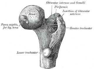
A femoral head ostectomy is a surgical operation to remove the head and neck from the femur. It is performed to alleviate pain, and is a salvage procedure, reserved for condition where pain can not be alleviated in any other way. It is common in veterinary surgery. Other names are excision arthroplasty of the femoral head and neck, Girdlestone's operation, Girdlestone procedure, and femoral head and neck ostectomy.

The Ortolani test is part of the physical examination for developmental dysplasia of the hip, along with the Barlow maneuver. Specifically, the Ortolani test is positive when a posterior dislocation of the hip is reducible with this maneuver. This is part of the standard infant exam performed preferably in early infancy.The Ortolani test is named after Marino Ortolani, who developed it in 1937.

Hip dysplasia is an abnormality of the hip joint where the socket portion does not fully cover the ball portion, resulting in an increased risk for joint dislocation. Hip dysplasia may occur at birth or develop in early life. Regardless, it does not typically produce symptoms in babies less than a year old. Occasionally one leg may be shorter than the other. The left hip is more often affected than the right. Complications without treatment can include arthritis, limping, and low back pain.