
In human anatomy, the arm is the part of the upper limb between the glenohumeral joint and the elbow joint. In common usage, the arm extends to the hand. It can be divided into the upper arm, which extends from the shoulder to the elbow, the forearm which extends from the elbow to the hand, and the hand. Anatomically the shoulder girdle with bones and corresponding muscles is by definition a part of the arm. The Latin term brachium may refer to either the arm as a whole or to the upper arm on its own.

The ulna is a long bone found in the forearm that stretches from the elbow to the smallest finger, and when in anatomical position, is found on the medial side of the forearm. It runs parallel to the radius, the other long bone in the forearm, and is the larger and longer of the two.
The forearm is the region of the upper limb between the elbow and the wrist. The term forearm is used in anatomy to distinguish it from the arm, a word which is most often used to describe the entire appendage of the upper limb, but which in anatomy, technically, means only the region of the upper arm, whereas the lower "arm" is called the forearm. It is homologous to the region of the leg that lies between the knee and the ankle joints, the crus.

The radius or radial bone is one of the two large bones of the forearm, the other being the ulna. It extends from the lateral side of the elbow to the thumb side of the wrist and runs parallel to the ulna. The radius is shorter and smaller than the ulna. It is a long bone, prism-shaped and slightly curved longitudinally.

A bone fracture is a medical condition in which there is a partial or complete break in the continuity of the bone. In more severe cases, the bone may be broken into several pieces. A bone fracture may be the result of high force impact or stress, or a minimal trauma injury as a result of certain medical conditions that weaken the bones, such as osteoporosis, osteopenia, bone cancer, or osteogenesis imperfecta, where the fracture is then properly termed a pathologic fracture.

An avulsion fracture is a bone fracture which occurs when a fragment of bone tears away from the main mass of bone as a result of physical trauma. This can occur at the ligament due to the application forces external to the body or at the tendon due to a muscular contraction that is stronger than the forces holding the bone together. Generally muscular avulsion is prevented due to the neurological limitations placed on muscle contractions. Highly trained athletes can overcome this neurological inhibition of strength and produce a much greater force output capable of breaking or avulsing a bone.

The upper or proximal extremity of the humerus consists of the bone's large rounded head joined to the body by a constricted portion called the neck, and two eminences, the greater and lesser tubercles.

The surgical neck of the humerus is a constriction below the tubercles of the greater tubercle and lesser tubercle, and above the Deltoid Tuberosity.

The humeroradial joint is the joint between the head of the radius and the capitulum of the humerus, is a limited ball-and-socket joint, hinge type of synovial joint.

Cubitus varus is a common deformity in which the extended forearm is deviated towards midline of the body.
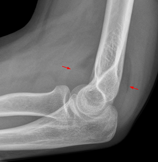
The fat pad sign, also known as the sail sign, is a potential finding on elbow radiography which suggests a fracture of one or more bones at the elbow. It is may indicate an occult fracture that is not directly visible. Its name derives from the fact that it has the shape of a spinnaker (sail). It is caused by displacement of the fat pad around the elbow joint. Both anterior and posterior fat pad signs exist, and both can be found on the same X-ray.

The elbow is the visible joint between the upper and lower parts of the arm. It includes prominent landmarks such as the olecranon, the elbow pit, the lateral and medial epicondyles, and the elbow joint. The elbow joint is the synovial hinge joint between the humerus in the upper arm and the radius and ulna in the forearm which allows the forearm and hand to be moved towards and away from the body.

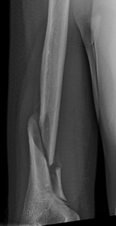
A supracondylar humerus fracture is a fracture of the distal humerus just above the elbow joint. The fracture is usually transverse or oblique and above the medial and lateral condyles and epicondyles. This fracture pattern is relatively rare in adults, but is the most common type of elbow fracture in children. In children, many of these fractures are non-displaced and can be treated with casting. Some are angulated or displaced and are best treated with surgery. In children, most of these fractures can be treated effectively with expectation for full recovery. Some of these injuries can be complicated by poor healing or by associated blood vessel or nerve injuries with serious complications.
A Holstein–Lewis fracture is a fracture of the distal third of the humerus resulting in entrapment of the radial nerve.
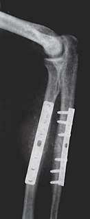
Internal fixation is an operation in orthopedics that involves the surgical implementation of implants for the purpose of repairing a bone, a concept that dates to the mid-nineteenth century and was made applicable for routine treatment in the mid-twentieth century. An internal fixator may be made of stainless steel, titanium alloy, or cobalt-chrome alloy.
The Milch classification is a system of categorizing single column distal humerus fractures based on the pattern of epicondyle involvement. It is distinct from the Jupiter classification which is used for bicolumnar distal humerus fractures.
The Gartland classification is a system of categorizing supracondylar humerus fractures, clinically useful as it predicts the likelihood of associated neurovascular injury, such as anterior interosseous nerve neurapraxia or brachial artery disruption.

A proximal humerus fracture is a break of the upper part of the bone of the arm (humerus). Symptoms include pain, swelling, and a decreased ability to move the shoulder. Complications may include axillary nerve or axillary artery injury.

