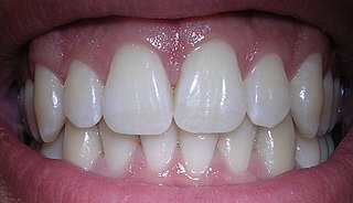
Human teeth function to mechanically break down items of food by cutting and crushing them in preparation for swallowing and digesting. As such, they are considered part of the human digestive system. Humans have four types of teeth: incisors, canines, premolars, and molars, which each have a specific function. The incisors cut the food, the canines tear the food and the molars and premolars crush the food. The roots of teeth are embedded in the maxilla or the mandible and are covered by gums. Teeth are made of multiple tissues of varying density and hardness.

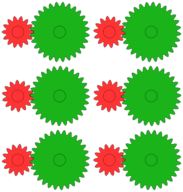
A gear is a rotating circular machine part having cut teeth or, in the case of a cogwheel or gearwheel, inserted teeth, which mesh with another (compatible) toothed part to transmit rotational power. While doing so, they can change the torque and rotational speed being transmitted and also change the rotational axis of the power being transmitted. The teeth on the two meshing gears all have the same shape.

In mammalian oral anatomy, the canine teeth, also called cuspids, dogteeth, eye teeth, vampire teeth, or vampire fangs, are the relatively long, pointed teeth. In the context of the upper jaw, they are also known as fangs. They can appear more flattened, however, causing them to resemble incisors and leading them to be called incisiform. They developed and are used primarily for firmly holding food in order to tear it apart, and occasionally as weapons. They are often the largest teeth in a mammal's mouth. Individuals of most species that develop them normally have four, two in the upper jaw and two in the lower, separated within each jaw by incisors; humans and dogs are examples. In most species, canines are the anterior-most teeth in the maxillary bone. The four canines in humans are the two upper maxillary canines and the two lower mandibular canines. They are specially prominent in dogs (Canidae), hence the name.

The Siegfried Line, known in German as the Westwall, was a German defensive line built during the late 1930s. Started in 1936, opposite the French Maginot Line, it stretched more than 630 km (390 mi) from Kleve on the border with the Netherlands, along the western border of Nazi Germany, to the town of Weil am Rhein on the border with Switzerland. The line featured more than 18,000 bunkers, tunnels and tank traps.

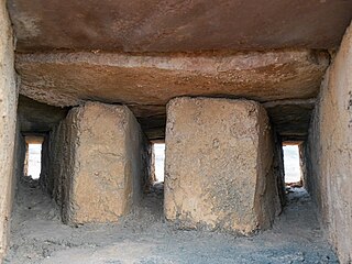
An embrasure is the opening in a battlement between two raised solid portions (merlons). Alternatively, an embrasure can be a space hollowed out throughout the thickness of a wall by the establishment of a bay. This term designates the internal part of this space, relative to the closing device, door or window. In fortification this refers to the outward splay of a window or of an arrowslit on the inside.

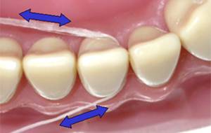
Dental floss is a cord of thin filaments used in interdental cleaning to remove food and dental plaque from between teeth or places a toothbrush has difficulty reaching or is unable to reach. Its regular use as part of oral cleaning is intended to maintain oral health.

The gums or gingiva consist of the mucosal tissue that lies over the mandible and maxilla inside the mouth. Gum health and disease can have an effect on general health.

Acid erosion is a type of tooth wear. It is defined as the irreversible loss of tooth structure due to chemical dissolution by acids not of bacterial origin. Dental erosion is the most common chronic condition of children ages 5–17, although it is only relatively recently that it has been recognised as a dental health problem. There is widespread ignorance of the damaging effects of acid erosion; this is particularly the case with erosion due to consumption of fruit juices because they tend to be seen as healthy. Acid erosion begins initially in the enamel, causing it to become thin, and can progress into dentin, giving the tooth a dull yellow appearance and leading to dentin hypersensitivity.

The mandibular central incisor is the tooth located on the jaw, adjacent to the midline of the face. It is mesial from both mandibular lateral incisors. As with all incisors, its function includes shearing or cutting food during mastication, commonly known as chewing. There are no cusps on the tooth. Instead, the surface area of the tooth used in eating is called an incisal ridge or incisal edge. Though the two are similar, there are some minor differences between the deciduous (baby) mandibular central incisor and that of the permanent mandibular central incisor. The mandibular central incisors are usually the first teeth to appear in the mouth, typically around the age of 6–8 months.

Pericoronitis is inflammation of the soft tissues surrounding the crown of a partially erupted tooth, including the gingiva (gums) and the dental follicle. The soft tissue covering a partially erupted tooth is known as an operculum, an area which can be difficult to access with normal oral hygiene methods. The hyponym operculitis technically refers to inflammation of the operculum alone.

The mandibular second premolar is the tooth located distally from both the mandibular first premolars of the mouth but mesial from both mandibular first molars. The function of this premolar is assist the mandibular first molar during mastication, commonly known as chewing. Mandibular second premolars have three cusps. There is one large cusp on the buccal side of the tooth. The lingual cusps are well developed and functional. Therefore, whereas the mandibular first premolar resembles a small canine, the mandibular second premolar is more alike to the first molar. There are no deciduous (baby) mandibular premolars. Instead, the teeth that precede the permanent mandibular premolars are the deciduous mandibular molars.

Toothlessness or edentulism is the condition of having no teeth. In organisms that naturally have teeth, it is the result of tooth loss. Organisms that never possessed teeth can also be described as edentulous. Examples are the members of the former zoological classification order of Edentata, which included anteaters and sloths, as they possess no anterior teeth and no or poorly developed posterior teeth.
Dental anatomy is a field of anatomy dedicated to the study of human tooth structures. The development, appearance, and classification of teeth fall within its purview. Tooth formation begins before birth, and the teeth's eventual morphology is dictated during this time. Dental anatomy is also a taxonomical science: it is concerned with the naming of teeth and the structures of which they are made, this information serving a practical purpose in dental treatment.
This is a list of definitions of commonly used terms of location and direction in dentistry. This set of terms provides orientation within the oral cavity, much as anatomical terms of location provide orientation throughout the body.
The gingival fibers are the connective tissue fibers that inhabit the gingival tissue adjacent to teeth and help hold the tissue firmly against the teeth. They are primarily composed of type I collagen, although type III fibers are also involved.
The interdental papilla, also known as the interdental gingiva, is the part of the gums (gingiva) that exists coronal to the free gingival margin on the mesial and distal surfaces of the teeth. The interdental papillae fill in the area between the teeth apical to their contact areas to prevent food impaction; they assume a conical shape for the anterior teeth and a blunted shape buccolingually for the posterior teeth.

An impacted tooth is one that fails to erupt into the dental arch within the expected developmental window. Because impacted teeth do not erupt, they are retained throughout the individual's lifetime unless extracted or exposed surgically. Teeth may become impacted because of adjacent teeth, dense overlying bone, excessive soft tissue or a genetic abnormality. Most often, the cause of impaction is inadequate arch length and space in which to erupt. That is the total length of the alveolar arch is smaller than the tooth arch. The wisdom teeth are frequently impacted because they are the last teeth to erupt in the oral cavity. Mandibular third molars are more commonly impacted than their maxillary counterparts.

An open contact is a term used in dentistry to describe the space between adjacent teeth when the teeth are neither touching nor a sufficient distance from each other to potentially allow the space to naturally remain free of debris.
Height of curvature in the tooth can be defined as the line encircling a tooth at its greatest bulge to a selected path of insertion. The height of curvature is the same as the height of contour.

Angularis nigra, Latin for 'black angle', also known as open gingival embrasures, and colloquially known as "black triangle", is the space or gap seen at the cervical embrasure, below the contact point of some teeth. The interdental papilla does not fully enclose the space, leading to an aperture between adjacent teeth. This gap has many causes including gingival recession, and gingival withdrawal post-orthodontic work. Interdental "black triangles" were rated as the third-most-disliked aesthetic problem below caries and crown margins. Treatment of angularis nigra often requires an interdisciplinary approach, involving periodontal, orthodontic and restorative treatment. Possible treatments to correct angularis nigra include addition of composite resin in the space, veneer placement, or gum graft. Angularis nigra is generally only treated based on the aesthetic preference of the patient.


