Related Research Articles

The human teeth function to mechanically break down items of food by cutting and crushing them in preparation for swallowing and digesting. Humans have four types of teeth: incisors, canines, premolars, and molars, which each have a specific function. The incisors cut the food, the canines tear the food and the molars and premolars crush the food. The roots of teeth are embedded in the maxilla or the mandible and are covered by gums. Teeth are made of multiple tissues of varying density and hardness.

Cementum is a specialized calcified substance covering the root of a tooth. The cementum is the part of the periodontium that attaches the teeth to the alveolar bone by anchoring the periodontal ligament.

Tooth enamel is one of the four major tissues that make up the tooth in humans and many other animals, including some species of fish. It makes up the normally visible part of the tooth, covering the crown. The other major tissues are dentin, cementum, and dental pulp. It is a very hard, white to off-white, highly mineralised substance that acts as a barrier to protect the tooth but can become susceptible to degradation, especially by acids from food and drink. Calcium hardens the tooth enamel. In rare circumstances enamel fails to form, leaving the underlying dentin exposed on the surface.

Tooth decay, also known as dental caries or cavities, is the breakdown of teeth due to acids made by bacteria. The cavities may be a number of different colors from yellow to black. Symptoms may include pain and difficulty with eating. Complications may include inflammation of the tissue around the tooth, tooth loss and infection or abscess formation.

Dentin or dentine is a calcified tissue of the body and, along with enamel, cementum, and pulp, is one of the four major components of teeth. It is usually covered by enamel on the crown and cementum on the root and surrounds the entire pulp. By volume, 45% of dentin consists of the mineral hydroxyapatite, 33% is organic material, and 22% is water. Yellow in appearance, it greatly affects the color of a tooth due to the translucency of enamel. Dentin, which is less mineralized and less brittle than enamel, is necessary for the support of enamel. Dentin rates approximately 3 on the Mohs scale of mineral hardness. There are two main characteristics which distinguish dentin from enamel: firstly, dentin forms throughout life; secondly, dentin is sensitive and can become hypersensitive to changes in temperature due to the sensory function of odontoblasts, especially when enamel recedes and dentin channels become exposed.

The pulp is the part in the centre of a tooth made up of living connective tissues and odontoblasts. The pulp is a part of the dentin–pulp complex (endodontium). The vitality of the dentin-pulp complex, both during health and after injury, depends on pulp cell activity and the signalling processes that regulate the cell's behaviour.

Ameloblasts are cells present only during tooth development that deposit tooth enamel, which is the hard outermost layer of the tooth forming the surface of the crown.
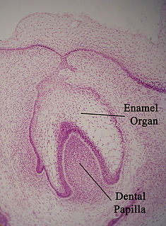
The enamel organ, also known as the dental organ, is a cellular aggregation seen in a developing tooth and it lies above the dental papilla. The enamel organ is responsible for the formation of enamel, initiation of dentine formation, establishment of the shape of a tooth's crown, and establishment of the dentoenamel junction.

Tooth development or odontogenesis is the complex process by which teeth form from embryonic cells, grow, and erupt into the mouth. For human teeth to have a healthy oral environment, all parts of the tooth must develop during appropriate stages of fetal development. Primary (baby) teeth start to form between the sixth and eighth week of prenatal development, and permanent teeth begin to form in the twentieth week. If teeth do not start to develop at or near these times, they will not develop at all, resulting in hypodontia or anodontia.
Amelogenesis is the formation of enamel on teeth and begins when the crown is forming during the advanced bell stage of tooth development after dentinogenesis forms a first layer of dentin. Dentin must be present for enamel to be formed. Ameloblasts must also be present for dentinogenesis to continue.

In embryology and prenatal development, the dental papilla is a condensation of ectomesenchymal cells called odontoblasts, seen in histologic sections of a developing tooth. It lies below a cellular aggregation known as the enamel organ. The dental papilla appears after 8–10 weeks intra uteral life. The dental papilla gives rise to the dentin and pulp of a tooth.
In vertebrates, an odontoblast is a cell of neural crest origin that is part of the outer surface of the dental pulp, and whose biological function is dentinogenesis, which is the formation of dentin, the substance beneath the tooth enamel on the crown and the cementum on the root.
Dentinogenesis is the formation of dentin, a substance that forms the majority of teeth. Dentinogenesis is performed by odontoblasts, which are a special type of biological cell on the outer wall of dental pulps, and it begins at the late bell stage of a tooth development. The different stages of dentin formation after differentiation of the cell result in different types of dentin: mantle dentin, primary dentin, secondary dentin, and tertiary dentin.

Burrs or burs are small cutting tools; not to be confused with small pieces of metal formed from cutting metal, used in die grinders, rotary tools, or dental drills. The name may be considered appropriate when their small-sized head is compared to a bur or their teeth are compared to a metal burr.

Polytrichum commune is a species of moss found in many regions with high humidity and rainfall. The species can be exceptionally tall for a moss with stems often exceeding 30 cm (12 in) though rarely reaching 70 cm (27.5 in), but it is most commonly found at shorter lengths of 5 to 10 cm. It is widely distributed throughout temperate and boreal latitudes in the Northern Hemisphere and also found in Mexico, several Pacific Islands including New Zealand, and also in Australia. It typically grows in bogs, wet heathland and along forest streams.

Enamel tufts are hypomineralized ribbon-like structures that run longitudinally to the tooth axis and extend from the dentinoenamel junction (DEJ) one fifth to a third into the enamel. They are called ‘‘tufts’’ due to their wavy look within the enamel microstructure.
Enamel lamellae are a type of hypomineralized structure in teeth that extend either from the dentinoenamel junction (DEJ) to the surface of the enamel, or vice versa. In essence, they are prominent linear enamel defects, but are of no clinical consequence. These structures contain proteins, proteoglycans, and lipids.
Dental pertains to the teeth, including dentistry. Topics related to the dentistry, the human mouth and teeth include:
Enamel hypocalcification is a defect of tooth enamel in which normal amounts of enamel are produced but are hypomineralized. In this defect the enamel is softer than normal. Some areas in enamel are hypocalcified: enamel spindles, enamel tufts, and enamel lamellae.

Enamelled glass or painted glass is glass which has been decorated with vitreous enamel and then fired to fuse the glasses. It can produce brilliant and long-lasting colours, and be translucent or opaque. Unlike most methods of decorating glass, it allows painting using several colours, and along with glass engraving, has historically been the main technique used to create the full range of image types on glass.
References
| | This dentistry article is a stub. You can help Wikipedia by expanding it. |