Enterobacterial common antigen

Last updated

The enterobacterial common antigen (ECA) is a carbohydrate antigen found in the outer membrane of many Enterobacterales species. [1] The antigen is unanimously absent from other gram-negative and gram-positive bacteria. [1] Aeromonas hydrophila 209A is the only organism outside of Enterobacterales that expresses the ECA. [1] More studies are needed to explain the presence of the antigen in this species as no other strains of this species express the antigen. The ECA is a polysaccharide made of repeating units of trisaccharides. The functions of these units have very few proven functions. Some evidence indicates role in pathogenicity in the bacteria that present the ECA. There are three separate types of ECA these include ECAPG, ECALPS, and ECACYC, each have different lengths. The synthesis of the ECA is controlled by the wec operon and has a 12-step synthesis which is described below. Due to the lack of proven function of the ECA, any clinical significance is hard to define however, some evidence suggests that human serum has antibodies against ECA.

Contents

History

The ECA was first described in 1962 in a paper written by Calvin M. Kunin and colleagues. [2] When documenting strains of E. coli responsible for urinary tract infections, Kunin exposed these E. coli strains to rabbit antisera and various other E. coli strains (102 homologous and heterologous strains).  Using passive agglutination, Kunin detected the O-antigen found in the lipopolysaccharide (LPS) of E. coli. During the experiments, Kunin noticed that there was cross-reactivity between the rabbit antisera and many of the E. coli strains. One of the sera, O14, reacted to an antigen found in a large range of E. coli strains. Notably, the antigen was not attached to the O-antigen of the LPS. The team noted that this antigen was observed in several other enteric (gram negative) bacteria strains and absent in many gram-positive strains. Kunin wanted to name the antigen the Common Antigen (CA) but his team convinced him to reconsider as to avoid confusion with the abbreviation for cancer. Thus, with a more specific name, the antigen was named the Enterobacterial Common Antigen (ECA). [2] [1] However, the ECA is not necessarily expressed in all enteric species. Many endosymbionts from Enterobacterales have lost the genes necessary for ECA synthesis. [1] Additionally, Aeromonas hydrophila 209A is the only organism outside of Enterobacterales that expresses the ECA. Yet, more studies are needed to explain the presence of the antigen in this species as no other strains of this species express the antigen. [1] Research on the ECA continues to develop.

Gene

The genes the regulate and control the synthesis of the ECA are located within on operon called the wec operon. The operon starts at 85.4 centisommes on the Escherichia coli K-12 chromosome. In regards to ECA, the wec operon has multiple genes that play important parts in inhibition as well as assembly of the ECA; these include but are not limited to wecB, wecC, wecD, wecE, wecA, and wecG. [1]

Structure

The polysaccharide of the enterobacterial common antigen (ECA) is composed of repeating units of a trisaccharide. This trisaccharide is made of N-acetylglucosamine (GlcNAc), N-Acetyl-D-Mannosaminuronic Acid (ManNAcA), and 4-acetamido-4,6-dideoxy-D-galactose (Fuc4NAc). GlcNAc is connected to ManNAcA via an alpha 1,4 linkage. ManNAcA is connected to Fuc4NAc via a beta 1,4 linkage. Each full trisaccharide unit is connected to each other by an alpha 1,3 linkage from Fuc4NAc to GlcNAc. [1] [3] [4]

There are three types of fully formed ECA: ECAPG, ECALPS, and ECACYC. [1] [5] [4] Regardless of the length, the polysaccharide is attached to a diacylglycerol with a phosphodiester bond and sits on the outer membrane of the bacterium. Like the ECAPG, the ECALPS sits on the outer membrane of the bacterium and is attached to a lipid. The ECALPS is not only attached to a lipid but also an LPS core. The final type of ECA is ECACYC, which is different from the other two types in that it is found only in the cytoplasm, and is composed of the polysaccharide bound in a ring. Another distinguishing characteristic of ECACYC is the total length of the trisaccharide chain; ECACYC is usually 4 to 6 units long, while ECALPS and ECAPG range from 1 to 14 units long. [6]

Synthesis

The three types of ECA have different qualities, but altogether share some basic features, the most important is the synthesis of the ECA unit. The synthesis of a unit of ECA is carried out by multiple enzymes. Each monosaccharide that makes up a unit is carried by an undecaprenyl phosphate (UDP) to a lipid carrier made of approximately 55 isoprenoid units in the inner membrane. Each monosaccharide is added to the lipid carrier to make a trisaccharide attached to a lipid. The enzymes that catalyze the group transfers of the monosaccharides from undecaprenyl phosphate to the forming ECA trisaccharide unit are Wec enzymes. The details of the synthesis are as follows: [1] [7] [6]

Step 1: WecA takes GlcNAc from UDP-GlcNAc and attaches GlcNAc-1-phosphate to an isoprenoid carrier. The product is called Lipid IECA.

Step 2: WecB takes UDP-GlcNAc and epimerizes it at carbon 2. This makes UDP-N-acetylmannosamine.

Step 3: WecC forms UDP-ManNAcA from the UDP-N-acetylmannosamine (from step 2) by reducing NAD+ to NADH.

Step 4: WecG takes the ManNAcA from UDP-ManNAcA (from step 3) and adds it to Lipid IECA (From step 1). The product of this group transfer is Lipid IIECA. At this point, 2 of the 3 carbohydrates that make the repeating unit of ECA are connected. The next steps (steps 5–8) prepare the final monosaccharide to be transferred to Lipid IIECA to make Lipid IIIECA, which has one full ECA trisaccharide unit.

Step 5: Glucose-1-phosphate, dTTP, and a H+ combine in a reaction catalyzed by RmIAECA to make dTDP-glucose.

Step 6: NAD+ acts as a cofactor for the enzyme RmIBECA to convert dTDP-glucose (from step 5) into dTDP-4-keto-6-deoxy-D-glucose. This happens through a series of redox reactions and a dehydration reaction.

Step 7: WecE adds an amino group from a glutamate residue to dTDP-4-keto-6-deoxy-D-glucose (from step 6) to make dTDP-4-amino-4,6-dideoxy-a-D-galactose (dTDP-Fuc4N)

Step 8: Acetyl-CoA acts as a cofactor for the enzyme WecD, which makes dTDP-Fuc4NAc acetylating dTDP-Fuc4N (from step 7).

Step 9: WecF takes Fuc4NAc from dTDP-Fuc4NAc (from step 8) and adds it to Lipid II (ECA), to make Lipid IIIECA. At this point, the complete trisaccharide unit is on the cytoplasmic side of the inner membrane attached to a lipid carrier (Lipid IIIECA). The final synthesis step, polymerization, occurs on the periplasmic side of the inner membrane.

Step 10: WzxE, a flippase, flips Lipid III from the cytoplasmic side of the inner membrane to the periplasmic side of the inner membrane.

Step 11: WzyE polymerizes the ECA trisaccharides by taking the ECA unit from the lipid carrier and WzxE. The lipid carrier is returned to the cytoplasmic side of the membrane to create another ECA trisaccharide unit.

Step 12: The growing ECA chain is stopped at the correct length by WzzE through an unknown mechanism. The product of these 12 steps is a long chain of ECA units attached to an isoprenoid lipid carrier. From here, the ECA polysaccharide is specialized into the type of completed ECA it will be.

To make ECAPG, the ECA polysaccharide is taken from the isoprenoid lipid carrier and given to a different, unknown lipid. The final product, ECAPG, is an ECA polymer linked by a phosphodiester bond to a diacylglycerol. [1]

To make ECALPS, WaaL, takes the ECA polysaccharide from the isoprenoid carrier and gives it to the core oligosaccharide of LPS. The synthesis of ECA in general shares many steps for the synthesis of LPS. The most significant overlap is the use of the WzxE and the WzzE proteins. [1]

The mechanism by which ECACYC is made is largely unknown. It has been previously established that ECACYC is synthesized in the periplasm and that this mechanism involves WzzE. From there, ECACYC is transported back across the inner membrane to the cytoplasm by a mechanism that has not yet been determined. [7]

Function

Due to the close association of ECA biosynthesis and other processes like O-antigen and peptidoglycan synthesis, it is difficult to define specific functions of the ECA in different species. [1] Enterobacterales species that have been cultured for prolonged periods show a significant reduction in O-chain synthesis while maintaining ECA stability. Several gene-knockout experiments show that upon altering genes participating in the synthesis of ECA, new sensitivities are observed. It is accepted that the ECA plays a role in pathogenicity. [1]

Clinical significance

The ECA occurs across all Enterobacterales species but very few species have antibodies to the antigen. This indicates that while all strains possess antigenic ECA, very few strains produce immunogenic ECA. [1]

Many studies reported low concentrations of ECA antibodies present in human serum occurred before ECA knockout strains were available. The reported values are a combined titer of the ECA antibodies as well as the antibodies that are bound to protein antigens that are shared among Enterobacterales. ECA antibodies have been detected in human serum after infection by E. coli, Yersinia enterocolitica O3 strains, or Proteus mirabilis-associated arthritis patients. [1]

Several studies have sought to identify how significant a role the ECA plays in pathogenicity, even under experimental conditions, the protection offered by an antibody response was slight and temporary in both active and passive immunization. [1] [4]

Related Research Articles

Peptidoglycan or murein is a unique large macromolecule, a polysaccharide, consisting of sugars and amino acids that forms a mesh-like peptidoglycan layer outside the plasma membrane, the rigid cell wall characteristic of most bacteria. The sugar component consists of alternating residues of β-(1,4) linked N-acetylglucosamine (NAG) and N-acetylmuramic acid (NAM). Attached to the N-acetylmuramic acid is an oligopeptide chain made of three to five amino acids. The peptide chain can be cross-linked to the peptide chain of another strand forming the 3D mesh-like layer. Peptidoglycan serves a structural role in the bacterial cell wall, giving structural strength, as well as counteracting the osmotic pressure of the cytoplasm. This repetitive linking results in a dense peptidoglycan layer which is critical for maintaining cell form and withstanding high osmotic pressures, and it is regularly replaced by peptidoglycan production. Peptidoglycan hydrolysis and synthesis are two processes that must occur in order for cells to grow and multiply, a technique carried out in three stages: clipping of current material, insertion of new material, and re-crosslinking of existing material to new material.

<span class="mw-page-title-main">Glycoprotein</span> Protein with oligosaccharide modifications

Glycoproteins are proteins which contain oligosaccharide chains covalently attached to amino acid side-chains. The carbohydrate is attached to the protein in a cotranslational or posttranslational modification. This process is known as glycosylation. Secreted extracellular proteins are often glycosylated.

<span class="mw-page-title-main">Teichoic acid</span>

Teichoic acids are bacterial copolymers of glycerol phosphate or ribitol phosphate and carbohydrates linked via phosphodiester bonds.

<span class="mw-page-title-main">Lipopolysaccharide</span> Class of molecules found in the outer membrane of Gram-negative bacteria

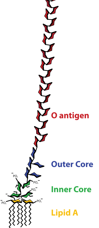
Lipopolysaccharides (LPS) are large molecules consisting of a lipid and a polysaccharide that are bacterial toxins. They are composed of an O-antigen, an outer core, and an inner core all joined by covalent bonds, and are found in the outer membrane of Gram-negative bacteria. Today, the term endotoxin is often used synonymously with LPS, although there are a few endotoxins that are not related to LPS, such as the so-called delta endotoxin proteins produced by Bacillus thuringiensis.

Dolichol refers to any of a group of long-chain mostly unsaturated organic compounds that are made up of varying numbers of isoprene units terminating in an α-saturated isoprenoid group, containing an alcohol functional group.

<span class="mw-page-title-main">Bacterial outer membrane</span>

The bacterial outer membrane is found in gram-negative bacteria. Its composition is distinct from that of the inner cytoplasmic cell membrane - among other things, the outer leaflet of the outer membrane of many gram-negative bacteria includes a complex lipopolysaccharide whose lipid portion acts as an endotoxin - and in some bacteria such as E. coli it is linked to the cell's peptidoglycan by Braun's lipoprotein.

<span class="mw-page-title-main">Glycosyltransferase</span> Class of enzymes that catalyze the transfer of glycosyl groups to an acceptor

Glycosyltransferases are enzymes that establish natural glycosidic linkages. They catalyze the transfer of saccharide moieties from an activated nucleotide sugar to a nucleophilic glycosyl acceptor molecule, the nucleophile of which can be oxygen- carbon-, nitrogen-, or sulfur-based.

<span class="mw-page-title-main">Tunicamycin</span> Chemical compound

Tunicamycin is a mixture of homologous nucleoside antibiotics that inhibits the UDP-HexNAc: polyprenol-P HexNAc-1-P family of enzymes. In eukaryotes, this includes the enzyme GlcNAc phosphotransferase (GPT), which catalyzes the transfer of N-acetylglucosamine-1-phosphate from UDP-N-acetylglucosamine to dolichol phosphate in the first step of glycoprotein synthesis. Tunicamycin blocks N-linked glycosylation (N-glycans) and treatment of cultured human cells with tunicamycin causes cell cycle arrest in G1 phase. It is used as an experimental tool in biology, e.g. to induce unfolded protein response. Tunicamycin is produced by several bacteria, including Streptomyces clavuligerus and Streptomyces lysosuperificus.

<span class="mw-page-title-main">UDP-glucose 4-epimerase</span> Class of enzymes

The enzyme UDP-glucose 4-epimerase, also known as UDP-galactose 4-epimerase or GALE, is a homodimeric epimerase found in bacterial, fungal, plant, and mammalian cells. This enzyme performs the final step in the Leloir pathway of galactose metabolism, catalyzing the reversible conversion of UDP-galactose to UDP-glucose. GALE tightly binds nicotinamide adenine dinucleotide (NAD+), a co-factor required for catalytic activity.

Nucleotide sugars are the activated forms of monosaccharides. Nucleotide sugars act as glycosyl donors in glycosylation reactions. Those reactions are catalyzed by a group of enzymes called glycosyltransferases.

In enzymology, a lipopolysaccharide N-acetylmannosaminouronosyltransferase is an enzyme that catalyzes the chemical reaction

In enzymology, a N-acetyllactosaminide 3-alpha-galactosyltransferase is an enzyme that catalyzes the chemical reaction

<span class="mw-page-title-main">DPAGT1</span> Protein-coding gene in the species Homo sapiens

UDP-N-acetylglucosamine—dolichyl-phosphate N-acetylglucosaminephosphotransferase is an enzyme that in humans is encoded by the DPAGT1 gene.

DTDP-4-amino-4,6-dideoxy-D-galactose acyltransferase is an enzyme with systematic name acetyl-CoA:dTDP-4-amino-4,6-dideoxy-alpha-D-galactose N-acetyltransferase. This enzyme catalyses the following chemical reaction

D-Man-alpha-(1->3) -D-Glc-beta-(1->4) -D-Glc-alpha-1-diphosphoundecaprenol 2-beta-glucuronyltransferase is an enzyme with the systematic name UDP-glucuronate: D-Man-alpha-(1->3) -D-Glc-beta-(1->4)-D-Glc-alpha-1-diphospho-ditrans,octacis-undecaprenol beta-1,2-glucuronyltransferase. This enzyme catalyses the following chemical reaction:

UDP-N-acetylglucosamine—undecaprenyl-phosphate N-acetylglucosaminephosphotransferase is an enzyme with systematic name UDP-N-acetyl-alpha-D-glucosamine:ditrans,octacis-undecaprenyl phosphate N-acetyl-alpha-D-glucosaminephosphotransferase. This enzyme catalyses the following chemical reaction

<span class="mw-page-title-main">Lipid II</span> Chemical compound

Lipid II is a precursor molecule in the synthesis of the cell wall of bacteria. It is a peptidoglycan, which is amphipathic and named for its bactoprenol hydrocarbon chain, which acts as a lipid anchor, embedding itself in the bacterial cell membrane. Lipid II must translocate across the cell membrane to deliver and incorporate its disaccharide-pentapeptide "building block" into the peptidoglycan mesh. Lipid II is the target of several antibiotics.

The multidrug/oligosaccharidyl-lipid/polysaccharide (MOP) flippase superfamily is a group of integral membrane protein families. The MOP flippase superfamily includes twelve distantly related families, six for which functional data are available:

  1. One ubiquitous family (MATE) specific for drugs - (TC# 2.A.66.1) The Multi Antimicrobial Extrusion (MATE) Family
  2. One (PST) specific for polysaccharides and/or their lipid-linked precursors in prokaryotes - (TC# 2.A.66.2) The Polysaccharide Transport (PST) Family
  3. One (OLF) specific for lipid-linked oligosaccharide precursors of glycoproteins in eukaryotes - (TC# 2.A.66.3) The Oligosaccharidyl-lipid Flippase (OLF) Family
  4. One (MVF) lipid-peptidoglycan precursor flippase involved in cell wall biosynthesis - (TC# 2.A.66.4) The Mouse Virulence Factor (MVF) Family
  5. One (AgnG) which includes a single functionally characterized member that extrudes the antibiotic, Agrocin 84 - (TC# 2.A.66.5) The Agrocin 84 Antibiotic Exporter (AgnG) Family
  6. And finally, one (Ank) that shuttles inorganic pyrophosphate (PPi) - (TC# 2.A.66.9) The Progressive Ankylosis (Ank) Family
<span class="mw-page-title-main">Methyltransferase/kinase WbdD</span>

Methyltransferase/kinase WbdD EC 2.1.1.294 and EC 2.7.1.181WbdD is a bifunctional enzyme that regulates the length of the LPS O-antigen polysaccharide chain. Stops the polymerization of the chain by phosphorylating and then methylating the phosphate on the terminal sugar. This terminal modification is essential for export of the O-antigen across the inner membrane. WbdD is also required for correct localization of the WbdA mannosyltransferase.

Undecaprenyl phosphate (UP), also known lipid-P, bactoprenol and C55-P., is a molecule with the primary function of trafficking polysaccharides across the cell membrane, largely contributing to the overall structure of the cell wall in Gram-positive bacteria. In some situations, UP can also be utilized to carry other cell-wall polysaccharides, but UP is the designated lipid carrier for peptidoglycan. During the process of carrying the peptidoglycan across the cell membrane, N-acetylglucosamine and N-acetylmuramic acid are linked to UP on the cytoplasmic side of the membrane before being carried across. UP works in a cycle of phosphorylation and dephosphorylation as the lipid carrier gets used, recycled, and reacts with undecaprenyl phosphate. This type of synthesis is referred to as de novo synthesis where a complex molecule is created from simpler molecules as opposed to a complete recycle of the entire structure.

References

  1. 1 2 3 4 5 6 7 8 9 10 11 12 13 14 15 16 17 Rai, A. K., & Mitchell, A. M. (2020). Enterobacterial Common Antigen: Synthesis and Function of an Enigmatic Molecule. MBio, 11(4), e01914-20. https://doi.org/10.1128/mBio.01914-20
  2. 1 2 Kunin, C. M.; Beard, M. V.; Halmagyi, N. E. (962). "Evidence for a Common Hapten Associated with Endotoxin Fractions of E. coli and other Enterobacteriaceae". Experimental Biology and Medicine. 111 (1): 160–166. doi:10.3181/00379727-111-27734. S2CID   86972326.
  3. Rahman, A., Barr, K., & Rick, P. D. (2001). Identification of the Structural Gene for the TDP-Fuc4NAc:Lipid II Fuc4NAc Transferase Involved in Synthesis of Enterobacterial Common Antigen in Escherichia coli K-12. Journal of Bacteriology, 183(22), 6509–6516. https://doi.org/10.1128/JB.183.22.6509-6516.2001
  4. 1 2 3 Kuhn, H.-M., Meier-Dieter, U., & Mayer, H. (1988). ECA, the enterobacterial common antigen. FEMS Microbiology Letters, 54(3), 195–222.
  5. Castelli, M. E., & Véscovi, E. G. (2011). The Rcs Signal Transduction Pathway Is Triggered by Enterobacterial Common Antigen Structure Alterations in Serratia marcescens. Journal of Bacteriology, 193(1), 63–74. https://doi.org/10.1128/JB.00839-10
  6. 1 2 Shen, X., Yang, Y., Li, P., Luo, H., & Kong, Q. (2021). Advances in the Research of Enterobacterial Common Antigen. Chinese Journal of Biotechnology, 37(4), 1081–1091. https://doi.org/10.13345/j.cjb.200334
  7. 1 2 Kajimura, J., Rahman, A., & Rick, P. D. (2005). Assembly of Cyclic Enterobacterial Common Antigen in Escherichia coli K-12. Journal of Bacteriology, 187(20), 6917–6927. https://doi.org/10.1128/JB.187.20.6917-6927.2005