The Erwin L. Hahn Institute for Magnetic Resonance Imaging [1] is a research institute which develops magnetic resonance imaging (MRI) for both cognitive neuroscience and medical diagnosis and treatment.
The Erwin L. Hahn Institute for Magnetic Resonance Imaging [1] is a research institute which develops magnetic resonance imaging (MRI) for both cognitive neuroscience and medical diagnosis and treatment.
It was founded in July 2005 by the University Duisburg-Essen (Germany) and the Radboud University Nijmegen (The Netherlands).
It is located on the grounds of the UNESCO World Cultural Heritage Zollverein in the Northeast of the city of Essen, Germany. The industrial complex Zollverein is a former coal mine and coke processing plant which represents an industry which dominated the industrialization of the Ruhr area in Germany during the 19th and 20th centuries.
The institute is named after Erwin L. Hahn, a physicist who has made innumerable contributions to the field of magnetic resonance. The centerpiece of the institute is a 7 Tesla whole-body magnet resonance imager from Siemens Healthcare, Erlangen, Germany. In contrast to the magnetic resonance imagers used in hospitals and clinics throughout the world, which commonly operate at a magnetic field strength of 1.5 Tesla, the ultra high magnetic field strength of this imager provides significantly higher sensitivity for structural and functional measurements of the human body.
The gauss, is a unit of measurement of magnetic induction, also known as magnetic flux density. The unit is part of the Gaussian system of units, which inherited it from the older centimetre–gram–second electromagnetic units (CGS-EMU) system. It was named after the German mathematician and physicist Carl Friedrich Gauss in 1936. One gauss is defined as one maxwell per square centimetre.

Magnetic resonance imaging (MRI) is a medical imaging technique used in radiology to form pictures of the anatomy and the physiological processes of the body. MRI scanners use strong magnetic fields, magnetic field gradients, and radio waves to generate images of the organs in the body. MRI does not involve X-rays or the use of ionizing radiation, which distinguishes it from computed tomography (CT) and positron emission tomography (PET) scans. MRI is a medical application of nuclear magnetic resonance (NMR) which can also be used for imaging in other NMR applications, such as NMR spectroscopy.

A Tesla coil is an electrical resonant transformer circuit designed by inventor Nikola Tesla in 1891. It is used to produce high-voltage, low-current, high-frequency alternating-current electricity. Tesla experimented with a number of different configurations consisting of two, or sometimes three, coupled resonant electric circuits.
Technological applications of superconductivity include:
The tesla is the unit of magnetic flux density in the International System of Units (SI).
Raymond Vahan Damadian was an Armenian-American physician, medical practitioner, and inventor of the first NMR scanning machine.

Electron paramagnetic resonance (EPR) or electron spin resonance (ESR) spectroscopy is a method for studying materials that have unpaired electrons. The basic concepts of EPR are analogous to those of nuclear magnetic resonance (NMR), but the spins excited are those of the electrons instead of the atomic nuclei. EPR spectroscopy is particularly useful for studying metal complexes and organic radicals. EPR was first observed in Kazan State University by Soviet physicist Yevgeny Zavoisky in 1944, and was developed independently at the same time by Brebis Bleaney at the University of Oxford.

The National High Magnetic Field Laboratory (MagLab) is a facility at Florida State University, the University of Florida, and Los Alamos National Laboratory in New Mexico, that performs magnetic field research in physics, biology, bioengineering, chemistry, geochemistry, biochemistry. It is the only such facility in the US, and is among twelve high magnetic facilities worldwide. The lab is supported by the National Science Foundation and the state of Florida, and works in collaboration with private industry.
Graham Wiggins was an American musician and scientist. He played the didgeridoo, keyboards, melodica, sampler, and various percussion instruments with his groups, the Oxford-based Outback and Dr. Didg. He also developed new technologies for magnetic resonance imaging (MRI).

Erwin Louis Hahn was an American physicist, best known for his work on nuclear magnetic resonance (NMR). In 1950 he discovered the spin echo.

Alexander Pines is an American chemist. He is the Glenn T. Seaborg Professor Emeritus, University of California, Berkeley, Chancellor's Professor Emeritus and Professor of the Graduate School, University of California, Berkeley, and a member of the California Institute for Quantitative Biosciences (QB3) and the Department of Bioengineering. He was born in 1945, grew up in Bulawayo in Southern Rhodesia and studied undergraduate mathematics and chemistry in Israel at Hebrew University of Jerusalem. Coming to the United States in 1968, Pines obtained his Ph.D. in chemical physics at M.I.T. in 1972 and joined the UC Berkeley faculty later that year.

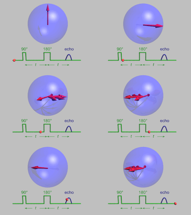
In magnetic resonance, a spin echo or Hahn echo is the refocusing of spin magnetisation by a pulse of resonant electromagnetic radiation. Modern nuclear magnetic resonance (NMR) and magnetic resonance imaging (MRI) make use of this effect.
Magnetic resonance can mean:

Nuclear magnetic resonance (NMR) is a physical phenomenon in which nuclei in a strong constant magnetic field are perturbed by a weak oscillating magnetic field and respond by producing an electromagnetic signal with a frequency characteristic of the magnetic field at the nucleus. This process occurs near resonance, when the oscillation frequency matches the intrinsic frequency of the nuclei, which depends on the strength of the static magnetic field, the chemical environment, and the magnetic properties of the isotope involved; in practical applications with static magnetic fields up to ca. 20 tesla, the frequency is similar to VHF and UHF television broadcasts (60–1000 MHz). NMR results from specific magnetic properties of certain atomic nuclei. Nuclear magnetic resonance spectroscopy is widely used to determine the structure of organic molecules in solution and study molecular physics and crystals as well as non-crystalline materials. NMR is also routinely used in advanced medical imaging techniques, such as in magnetic resonance imaging (MRI).

The physics of magnetic resonance imaging (MRI) concerns fundamental physical considerations of MRI techniques and technological aspects of MRI devices. MRI is a medical imaging technique mostly used in radiology and nuclear medicine in order to investigate the anatomy and physiology of the body, and to detect pathologies including tumors, inflammation, neurological conditions such as stroke, disorders of muscles and joints, and abnormalities in the heart and blood vessels among others. Contrast agents may be injected intravenously or into a joint to enhance the image and facilitate diagnosis. Unlike CT and X-ray, MRI uses no ionizing radiation and is, therefore, a safe procedure suitable for diagnosis in children and repeated runs. Patients with specific non-ferromagnetic metal implants, cochlear implants, and cardiac pacemakers nowadays may also have an MRI in spite of effects of the strong magnetic fields. This does not apply on older devices, and details for medical professionals are provided by the device's manufacturer.
Robert Turner is a British neuroscientist, physicist, and social anthropologist. He has been a director and professor at the Max Planck Institute for Human Cognitive and Brain Sciences in Leipzig, Germany, and is an internationally recognized expert in brain physics and magnetic resonance imaging (MRI). Coils inside every MRI scanner owe their shape to his ideas.
James S. Hyde was an American biophysicist. He held the James S. Hyde chair in Biophysics at the Medical College of Wisconsin (MCW) where he specialized in magnetic resonance instrumentation and methodology development in two distinct areas: electron paramagnetic resonance (EPR) spectroscopy and magnetic resonance imaging (MRI). He is senior author of the widely cited 1995 paper by B.B. Biswal et al. reporting the discovery of resting state functional connectivity (fcMRI) in the human brain. He also served as Director of the National Biomedical EPR Center, a Research Resource supported by the National Institutes of Health. He was author of more than 400 peer-reviewed papers and review articles and held 35 U.S. Patents. He was recognized by Festschrifts in both EPR and fcMRI.
The Gas Dynamic Trap is a magnetic mirror machine being operated at the Budker Institute of Nuclear Physics in Akademgorodok, Russia.

Magnetic resonance imaging (MRI) is in general a safe technique, although injuries may occur as a result of failed safety procedures or human error. During the last 150 years, thousands of papers focusing on the effects or side effects of magnetic or radiofrequency fields have been published. They can be categorized as incidental and physiological. Contraindications to MRI include most cochlear implants and cardiac pacemakers, shrapnel and metallic foreign bodies in the eyes. The safety of MRI during the first trimester of pregnancy is uncertain, but it may be preferable to other options. Since MRI does not use any ionizing radiation, its use generally is favored in preference to CT when either modality could yield the same information.
The history of magnetic resonance imaging (MRI) includes the work of many researchers who contributed to the discovery of nuclear magnetic resonance (NMR) and described the underlying physics of magnetic resonance imaging, starting early in the twentieth century. MR imaging was invented by Paul C. Lauterbur who developed a mechanism to encode spatial information into an NMR signal using magnetic field gradients in September 1971; he published the theory behind it in March 1973.