Related Research Articles

Cerebrospinal fluid (CSF) is a clear, colorless body fluid found within the tissue that surrounds the brain and spinal cord of all vertebrates.

Hydrocephalus is a condition in which an accumulation of cerebrospinal fluid (CSF) occurs within the brain. This typically causes increased pressure inside the skull. Older people may have headaches, double vision, poor balance, urinary incontinence, personality changes, or mental impairment. In babies, it may be seen as a rapid increase in head size. Other symptoms may include vomiting, sleepiness, seizures, and downward pointing of the eyes.

Colpocephaly is a cephalic disorder involving the disproportionate enlargement of the occipital horns of the lateral ventricles and is usually diagnosed early after birth due to seizures. It is a nonspecific finding and is associated with multiple neurological syndromes, including agenesis of the corpus callosum, Chiari malformation, lissencephaly, and microcephaly. Although the exact cause of colpocephaly is not known yet, it is commonly believed to occur as a result of neuronal migration disorders during early brain development, intrauterine disturbances, perinatal injuries, and other central nervous system disorders. Individuals with colpocephaly have various degrees of motor disabilities, visual defects, spasticity, and moderate to severe intellectual disability. No specific treatment for colpocephaly exists, but patients may undergo certain treatments to improve their motor function or intellectual disability.
Porencephaly is an extremely rare cephalic disorder involving encephalomalacia. It is a neurological disorder of the central nervous system characterized by cysts or cavities within the cerebral hemisphere. Porencephaly was termed by Heschl in 1859 to describe a cavity in the human brain. Derived from Greek roots, the word porencephaly means 'holes in the brain'. The cysts and cavities are more likely to be the result of destructive (encephaloclastic) cause, but can also be from abnormal development (malformative), direct damage, inflammation, or hemorrhage. The cysts and cavities cause a wide range of physiological, physical, and neurological symptoms. Depending on the patient, this disorder may cause only minor neurological problems, without any disruption of intelligence, while others may be severely disabled or die before the second decade of their lives. However, this disorder is far more common within infants, and porencephaly can occur both before or after birth.

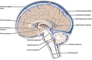
The ventricular system is a set of four interconnected cavities known as cerebral ventricles in the brain. Within each ventricle is a region of choroid plexus which produces the circulating cerebrospinal fluid (CSF). The ventricular system is continuous with the central canal of the spinal cord from the fourth ventricle, allowing for the flow of CSF to circulate.

The caudate nucleus is one of the structures that make up the corpus striatum, which is a component of the basal ganglia in the human brain. While the caudate nucleus has long been associated with motor processes due to its role in Parkinson's disease, it plays important roles in various other nonmotor functions as well, including procedural learning, associative learning and inhibitory control of action, among other functions. The caudate is also one of the brain structures which compose the reward system and functions as part of the cortico–basal ganglia–thalamic loop.

The choroid plexus, or plica choroidea, is a plexus of cells that arises from the tela choroidea in each of the ventricles of the brain. Regions of the choroid plexus produce and secrete most of the cerebrospinal fluid (CSF) of the central nervous system. The choroid plexus consists of modified ependymal cells surrounding a core of capillaries and loose connective tissue. Multiple cilia on the ependymal cells move to circulate the cerebrospinal fluid.

The fornix is a C-shaped bundle of nerve fibers in the brain that acts as the major output tract of the hippocampus. The fornix also carries some afferent fibers to the hippocampus from structures in the diencephalon and basal forebrain. The fornix is part of the limbic system. While its exact function and importance in the physiology of the brain are still not entirely clear, it has been demonstrated in humans that surgical transection—the cutting of the fornix along its body—can cause memory loss. There is some debate over what type of memory is affected by this damage, but it has been found to most closely correlate with recall memory rather than recognition memory. This means that damage to the fornix can cause difficulty in recalling long-term information such as details of past events, but it has little effect on the ability to recognize objects or familiar situations.

Cerebellar hypoplasia is characterized by reduced cerebellar volume, even though cerebellar shape is (near) normal. It consists of a heterogeneous group of disorders of cerebellar maldevelopment presenting as early-onset non–progressive congenital ataxia, hypotonia and motor learning disability.

Choroid plexus cysts (CPCs) are cysts that occur within choroid plexus of the brain. They are the most common type of intraventricular cyst, occurring in 1% of all pregnancies.

The lateral ventricles are the two largest ventricles of the brain and contain cerebrospinal fluid (CSF). Each cerebral hemisphere contains a lateral ventricle, known as the left or right lateral ventricle, respectively.

Periventricular leukomalacia (PVL) is a form of white-matter brain injury, characterized by the necrosis of white matter near the lateral ventricles. It can affect newborns and fetuses; premature infants are at the greatest risk of neonatal encephalopathy which may lead to this condition. Affected individuals generally exhibit motor control problems or other developmental delays, and they often develop cerebral palsy or epilepsy later in life. The white matter in preterm born children is particularly vulnerable during the third trimester of pregnancy when white matter developing takes place and the myelination process starts around 30 weeks of gestational age.

Arachnoid cysts are cerebrospinal fluid covered by arachnoidal cells and collagen that may develop between the surface of the brain and the cranial base or on the arachnoid membrane, one of the three meningeal layers that cover the brain and the spinal cord. Primary arachnoid cysts are a congenital disorder whereas secondary arachnoid cysts are the result of head injury or trauma. Most cases of primary cysts begin during infancy; however, onset may be delayed until adolescence.

Dandy–Walker malformation (DWM), also known as Dandy–Walker syndrome (DWS), is a rare congenital brain malformation in which the part joining the two hemispheres of the cerebellum does not fully form, and the fourth ventricle and space behind the cerebellum are enlarged with cerebrospinal fluid. Most of those affected develop hydrocephalus within the first year of life, which can present as increasing head size, vomiting, excessive sleepiness, irritability, downward deviation of the eyes and seizures. Other, less common symptoms are generally associated with comorbid genetic conditions and can include congenital heart defects, eye abnormalities, intellectual disability, congenital tumours, other brain defects such as agenesis of the corpus callosum, skeletal abnormalities, an occipital encephalocele or underdeveloped genitalia or kidneys. It is sometimes discovered in adolescents or adults due to mental health problems.
Akinetic mutism is a medical condition where patients tend neither to move (akinesia) nor speak (mutism). Akinetic mutism was first described in 1941 as a mental state where patients lack the ability to move or speak. However, their eyes may follow their observer or be diverted by sound. Patients lack most motor functions such as speech, facial expressions, and gestures, but demonstrate apparent alertness. They exhibit reduced activity and slowness, and can speak in whispered monosyllables. Patients often show visual fixation on their examiner, move their eyes in response to an auditory stimulus, or move after often repeated commands. Patients with akinetic mutism are not paralyzed, but lack the will to move. Many patients describe that as soon as they "will" or attempt a movement, a "counter-will" or "resistance" rises up to meet them.
Primitive reflexes are reflex actions originating in the central nervous system that are exhibited by normal infants, but not neurologically intact adults, in response to particular stimuli. These reflexes are suppressed by the development of the frontal lobes as a child transitions normally into child development. These primitive reflexes are also called infantile, infant or newborn reflexes.

A colloid cyst is a non-malignant tumor in the brain. It consists of a gelatinous material contained within a membrane of epithelial tissue. It is almost always found just posterior to the foramen of Monro in the anterior aspect of the third ventricle, originating from the roof of the ventricle. Because of its location, it can cause obstructive hydrocephalus and increased intracranial pressure. Colloid cysts represent 0.5–1.0% of intracranial tumors.

The cave of septum pellucidum (CSP), cavum septi pellucidi, or cavity of septum pellucidum is a slit-like space in the septum pellucidum that is present in fetuses but usually fuses during infancy. The septum pellucidum is a thin, laminated translucent vertical membrane in the midline of the brain separating the anterior horns of the right and left ventricles. It lies posterior to the corpus callosum. Persistence of the cave of septum pellucidum after infancy has been loosely associated with neural maldevelopment and several mental disorders that correlate with decreased brain tissue.

A cerebral shunt is a device permanently implanted inside the head and body to drain excess fluid away from the brain. They are commonly used to treat hydrocephalus, the swelling of the brain due to excess buildup of cerebrospinal fluid (CSF). If left unchecked, the excess CSF can lead to an increase in intracranial pressure (ICP), which can cause intracranial hematoma, cerebral edema, crushed brain tissue or herniation. The drainage provided by a shunt can alleviate or prevent these problems in patients with hydrocephalus or related diseases.
References
- ↑ Unger, S; Salem, S; Wylie, L; Shah, V (February 2011). "Newborn frontal horn cysts: cause for concern?". Journal of Perinatology. 31 (2): 98–103. doi:10.1038/jp.2010.79. PMID 20616785. S2CID 9691516.
- ↑ Sun, LR; Tekes, A; Golden, WC; Oakley, C (September 2014). "Connatal cyst in a preterm twin infant with maternal comorbidities". Pediatric Neurology. 51 (3): 444–7. doi:10.1016/j.pediatrneurol.2014.05.030. PMID 25023976.
- ↑ Chang, CL; Chiu, NC; Ho, CS; Li, ST (August 2006). "Frontal horn cysts in normal neonates". Brain & Development. 28 (7): 426–30. doi:10.1016/j.braindev.2006.01.002. PMC 7125929 . PMID 16503391.