Related Research Articles

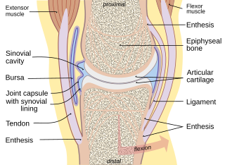
A joint or articulation is the connection made between bones, ossicles, or other hard structures in the body which link an animal's skeletal system into a functional whole. They are constructed to allow for different degrees and types of movement. Some joints, such as the knee, elbow, and shoulder, are self-lubricating, almost frictionless, and are able to withstand compression and maintain heavy loads while still executing smooth and precise movements. Other joints such as sutures between the bones of the skull permit very little movement in order to protect the brain and the sense organs. The connection between a tooth and the jawbone is also called a joint, and is described as a fibrous joint known as a gomphosis. Joints are classified both structurally and functionally.

The radius or radial bone is one of the two large bones of the forearm, the other being the ulna. It extends from the lateral side of the elbow to the thumb side of the wrist and runs parallel to the ulna. The ulna is longer than the radius, but the radius is thicker. The radius is a long bone, prism-shaped and slightly curved longitudinally.

A Colles' fracture is a type of fracture of the distal forearm in which the broken end of the radius is bent backwards. Symptoms may include pain, swelling, deformity, and bruising. Complications may include damage to the median nerve.

A bone fracture is a medical condition in which there is a partial or complete break in the continuity of any bone in the body. In more severe cases, the bone may be broken into several fragments, known as a comminuted fracture. An open fracture is a bone fracture where the broken bone breaks through the skin. A bone fracture may be the result of high force impact or stress, or a minimal trauma injury as a result of certain medical conditions that weaken the bones, such as osteoporosis, osteopenia, bone cancer, or osteogenesis imperfecta, where the fracture is then properly termed a pathologic fracture. Most bone fractures require urgent medical attention to prevent further injury.

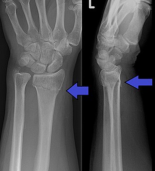
A distal radius fracture, also known as wrist fracture, is a break of the part of the radius bone which is close to the wrist. Symptoms include pain, bruising, and rapid-onset swelling. The ulna bone may also be broken.

A Smith's fracture, is a fracture of the distal radius.

A calcaneal fracture is a break of the calcaneus. Symptoms may include pain, bruising, trouble walking, and deformity of the heel. It may be associated with breaks of the hip or back.

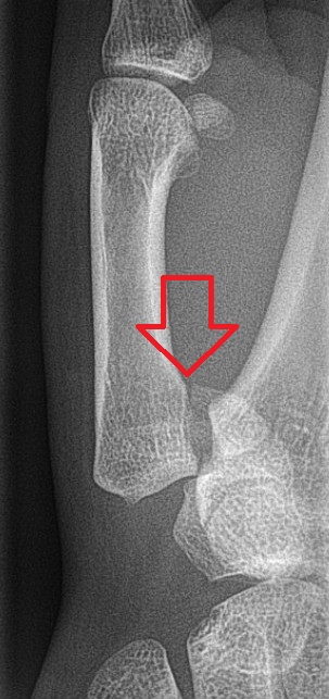
Bennett fracture is a type of partial broken finger involving the base of the thumb, and extends into the carpometacarpal (CMC) joint.

A Barton's fracture is a type of wrist injury where there is a broken bone associated with a dislocated bone in the wrist, typically occurring after falling on top of a bent wrist. It is an intra-articular fracture of the distal radius with dislocation of the radiocarpal joint.

A humerus fracture is a break of the humerus bone in the upper arm. Symptoms may include pain, swelling, and bruising. There may be a decreased ability to move the arm and the person may present holding their elbow. Complications may include injury to an artery or nerve, and compartment syndrome.

A supracondylar humerus fracture is a fracture of the distal humerus just above the elbow joint. The fracture is usually transverse or oblique and above the medial and lateral condyles and epicondyles. This fracture pattern is relatively rare in adults, but is the most common type of elbow fracture in children. In children, many of these fractures are non-displaced and can be treated with casting. Some are angulated or displaced and are best treated with surgery. In children, most of these fractures can be treated effectively with expectation for full recovery. Some of these injuries can be complicated by poor healing or by associated blood vessel or nerve injuries with serious complications.

A tibial plateau fracture is a break of the upper part of the tibia (shinbone) that involves the knee joint. This could involve the medial, lateral, central, or bicondylar. Symptoms include pain, swelling, and a decreased ability to move the knee. People are generally unable to walk. Complication may include injury to the artery or nerve, arthritis, and compartment syndrome.

Olecranon fracture is a fracture of the bony portion of the elbow. The injury is fairly common and often occurs following a fall or direct trauma to the elbow. The olecranon is the proximal extremity of the ulna which is articulated with the humerus bone and constitutes a part of the elbow articulation. Its location makes it vulnerable to direct trauma.
Frykman classification is a system of categorizing Colles' fractures. In the Frykman classification system there are four types of fractures.

A proximal humerus fracture is a break of the upper part of the bone of the arm (humerus). Symptoms include pain, swelling, and a decreased ability to move the shoulder. Complications may include axillary nerve or axillary artery injury.
Older's classification is a system of categorizing Colles' fractures, proposed in 1965. In the Older's classification system there are four types of fractures.
Lidström classification is a system of categorizing fractures of the distal radius, one of the two bones of the forearm. In the Lidström classification system there are six types of fractures. The classification system is based on fracture line, direction and degree of displacement, extent of articular involvement and involvement of the distal radioulnar joint, and was first published in 1959.
Nissen-Lie classification is a system of categorizing Colles' fractures. In the Nissen-Lie classification system there are seven types of fractures. The classification system was first published in 1939.

There are a number of ways to classify distal radius fractures. Classifications systems are devised to describe patterns of injury which will behave in predictable ways, to distinguish between conditions which have different outcomes or which need different treatments. Most wrist fracture systems have failed to accomplish any of these goals and there is no consensus about the most useful one.

A broken toe is a type of bone fracture. Symptoms include pain when the toe is touched near the break point, or compressed along its length. There may be bruising, swelling, stiffness, or displacement of the broken bone ends from their normal position.
References
- ↑ Berger, Richard A.; Weiss, Arnold-Peter C. (2004). Hand Surgery. Lippincott Williams & Wilkins. p. 249. ISBN 9780781728744 . Retrieved 13 November 2017.
- ↑ Fernandez, Diego L.; Jupiter, Jesse B. (2012). Fractures of the Distal Radius: A Practical Approach to Management. Springer Science & Business Media. p. 27. ISBN 9781468404784 . Retrieved 13 November 2017.