
Anatomy is the branch of biology concerned with the study of the structure of organisms and their parts. Anatomy is a branch of natural science that deals with the structural organization of living things. It is an old science, having its beginnings in prehistoric times. Anatomy is inherently tied to developmental biology, embryology, comparative anatomy, evolutionary biology, and phylogeny, as these are the processes by which anatomy is generated, both over immediate and long-term timescales. Anatomy and physiology, which study the structure and function of organisms and their parts respectively, make a natural pair of related disciplines, and are often studied together. Human anatomy is one of the essential basic sciences that are applied in medicine.

Andreas Vesalius was a 16th-century anatomist, physician, and author of one of the most influential books on human anatomy, De Humani Corporis Fabrica Libri Septem. Vesalius is often referred to as the founder of modern human anatomy. He was born in Brussels, which was then part of the Habsburg Netherlands. He was a professor at the University of Padua (1537–1542) and later became Imperial physician at the court of Emperor Charles V.
The history of anatomy extends from the earliest examinations of sacrificial victims to the sophisticated analyses of the body performed by modern anatomists and scientists. Written descriptions of human organs and parts can be traced back thousands of years to ancient Egyptian papyri, where attention to the body was necessitated by their highly elaborate burial practices.

The history of anatomy in the 19th century saw anatomists largely finalise and systematise the descriptive human anatomy of the previous century. The discipline also progressed to establish growing sources of knowledge in histology and developmental biology, not only of humans but also of animals.

William Hunter was a Scottish anatomist and physician. He was a leading teacher of anatomy, and the outstanding obstetrician of his day. His guidance and training of his equally famous brother, John Hunter, was also of great importance.

The accessory nerve, also known as the eleventh cranial nerve, cranial nerve XI, or simply CN XI, is a cranial nerve that supplies the sternocleidomastoid and trapezius muscles. It is classified as the eleventh of twelve pairs of cranial nerves because part of it was formerly believed to originate in the brain. The sternocleidomastoid muscle tilts and rotates the head, whereas the trapezius muscle, connecting to the scapula, acts to shrug the shoulder.

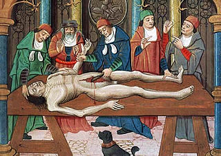
Dissection is the dismembering of the body of a deceased animal or plant to study its anatomical structure. Autopsy is used in pathology and forensic medicine to determine the cause of death in humans. Less extensive dissection of plants and smaller animals preserved in a formaldehyde solution is typically carried out or demonstrated in biology and natural science classes in middle school and high school, while extensive dissections of cadavers of adults and children, both fresh and preserved are carried out by medical students in medical schools as a part of the teaching in subjects such as anatomy, pathology and forensic medicine. Consequently, dissection is typically conducted in a morgue or in an anatomy lab.

Sir Richard Quain, 1st Baronet, was an Irish physician.

De Humani Corporis Fabrica Libri Septem is a set of books on human anatomy written by Andreas Vesalius (1514–1564) and published in 1543. It was a major advance in the history of anatomy over the long-dominant work of Galen, and presented itself as such.

Henry Gray was a British anatomist and surgeon most notable for publishing the book Gray's Anatomy. He was elected a Fellow of the Royal Society (FRS) at the age of 25.
Jones Quain was an Irish anatomist, born at Mallow. Quain was Professor of Anatomy and Physiology in the University of London. He was author of Elements of Anatomy, of which the first edition was published in 1828.

Govert Bidloo or Govard Bidloo was a Dutch Golden Age physician, anatomist, poet and playwright. He was the personal physician of William III of Orange-Nassau, Dutch stadholder and King of England, Scotland and Ireland.

Richard Quain was an English anatomist and surgeon, born at Fermoy, Ireland, a brother of Jones Quain. He studied medicine in London and in Paris. He was appointed demonstrator in 1828 and professor of anatomy in 1832 at the University of London, resigning in 1850, and assistant surgeon in 1834 and surgeon in 1848 to the North London Hospital, from which he resigned in 1866. He was president of the Royal College of Surgeons in 1868.

William Sharpey FRS FRSE LLD was a Scottish anatomist and physiologist. Sharpey became the outstanding exponent of experimental biology and is described as the "father of British physiology".
A cadaver or corpse is a dead human body that is used by medical students, physicians and other scientists to study anatomy, identify disease sites, determine causes of death, and provide tissue to repair a defect in a living human being. Students in medical school study and dissect cadavers as a part of their education. Others who study cadavers include archaeologists and arts students.

The Medical Renaissance, from around 1400 to 1700 CE, was a period of progress in European medical knowledge, with renewed interest in the ideas of the ancient Greek and Roman civilizations along with Arabic-Persian medicine, following the translation into Latin of many works from these societies. Medical discoveries during the Medical Renaissance are credited with paving the way for modern medicine.
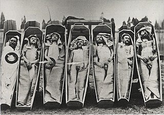
An anatomy murder is a murder committed in order to use all or part of the cadaver for medical research or teaching. It is not a medicine murder because the body parts are not believed to have any medicinal use in themselves. The motive for the murder is created by the demand for cadavers for dissection, and the opportunity to learn anatomy and physiology as a result of the dissection. Rumors concerning the prevalence of anatomy murders are associated with the rise in demand for cadavers in research and teaching produced by the Scientific Revolution. During the 19th century, the sensational serial murders associated with Burke and Hare and the London Burkers led to legislation which provided scientists and medical schools with legal ways of obtaining cadavers. Rumors persist that anatomy murders are carried out wherever there is a high demand for cadavers. These rumors, like those concerning organ theft, are hard to substantiate, and may reflect continued, deep-held fears of the use of cadavers as commodities.
John Barclay was a Scottish comparative anatomist, extramural teacher in anatomy, and director of the Highland Society of Scotland.

George Henry Ford aka G. H. Ford, was a South African natural history illustrator who joined the British Museum in 1837. He portrayed animals and produced the plates in Sir Andrew Smith's Illustrations of the Zoology of South Africa.
As anatomy classes in medical education proliferated in the 19th century, so too did the need for bodies to dissect. Grave robbery proliferated, along with associated social discontent, revulsion, and unhappiness. Conflicts arose between medical practitioners and defenders of bodies, graves and graveyards. This resulted in riots. Social legislation was passed in many countries to address the competing concerns.
