Related Research Articles

An axon, or nerve fiber, is a long, slender projection of a nerve cell, or neuron, in vertebrates, that typically conducts electrical impulses known as action potentials away from the nerve cell body. The function of the axon is to transmit information to different neurons, muscles, and glands. In certain sensory neurons, such as those for touch and warmth, the axons are called afferent nerve fibers and the electrical impulse travels along these from the periphery to the cell body and from the cell body to the spinal cord along another branch of the same axon. Axon dysfunction can be the cause of many inherited and acquired neurological disorders that affect both the peripheral and central neurons. Nerve fibers are classed into three types – group A nerve fibers, group B nerve fibers, and group C nerve fibers. Groups A and B are myelinated, and group C are unmyelinated. These groups include both sensory fibers and motor fibers. Another classification groups only the sensory fibers as Type I, Type II, Type III, and Type IV.

A nerve is an enclosed, cable-like bundle of nerve fibers in the peripheral nervous system.

Nervous tissue, also called neural tissue, is the main tissue component of the nervous system. The nervous system regulates and controls body functions and activity. It consists of two parts: the central nervous system (CNS) comprising the brain and spinal cord, and the peripheral nervous system (PNS) comprising the branching peripheral nerves. It is composed of neurons, also known as nerve cells, which receive and transmit impulses, and neuroglia, also known as glial cells or glia, which assist the propagation of the nerve impulse as well as provide nutrients to the neurons.

Afferent nerve fibers are axons of sensory neurons that carry sensory information from sensory receptors to the central nervous system. Many afferent projections arrive at a particular brain region.
Efferent nerve fibers refer to axonal projections that exit a particular region; as opposed to afferent projections that arrive at the region. These terms have a slightly different meaning in the context of the peripheral nervous system (PNS) and central nervous system (CNS). The efferent fiber is a long process projecting far from the neuron's body that carries nerve impulses away from the central nervous system toward the peripheral effector organs. A bundle of these fibers is called an efferent nerve. The opposite direction of neural activity is afferent conduction, which carries impulses by way of the afferent nerve fibers of sensory neurons.

A free nerve ending (FNE) or bare nerve ending, is an unspecialized, afferent nerve fiber sending its signal to a sensory neuron. Afferent in this case means bringing information from the body's periphery toward the brain. They function as cutaneous nociceptors and are essentially used by vertebrates to detect noxious stimuli that often result in pain.

The dorsal root of spinal nerve is one of two "roots" which emerge from the spinal cord. It emerges directly from the spinal cord, and travels to the dorsal root ganglion. Nerve fibres with the ventral root then combine to form a spinal nerve. The dorsal root transmits sensory information, forming the afferent sensory root of a spinal nerve.
The cochlear nerve is one of two parts of the vestibulocochlear nerve, a cranial nerve present in amniotes, the other part being the vestibular nerve. The cochlear nerve carries auditory sensory information from the cochlea of the inner ear directly to the brain. The other portion of the vestibulocochlear nerve is the vestibular nerve, which carries spatial orientation information to the brain from the semicircular canals, also known as semicircular ducts.
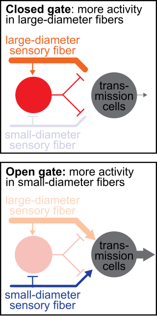
The gate control theory of pain asserts that non-painful input closes the nerve "gates" to painful input, which prevents pain sensation from traveling to the central nervous system.
A sensory nerve, or afferent nerve, is an anatomic term for a nerve which contains exclusively afferent nerve fibers. Nerves containing also motor fibers are called mixed. Afferent nerve fibers in a sensory nerve carry sensory information toward the central nervous system (CNS) from different sensory receptors of sensory neurons in the peripheral nervous system.

The white ramus communicans from Latin ramus (branch) and communicans (communicating) is the preganglionic sympathetic outflow nerve tract from the spinal cord.

The sympathetic ganglia, or paravertebral ganglia are autonomic ganglia, of the sympathetic nervous system. Ganglia are 20,000 to 30,000 afferent and efferent nerve cell bodies that run along on either side of the spinal cord. Afferent nerve cell bodies bring information from the body to the brain and spinal cord, while efferent nerve cell bodies bring information from the brain and spinal cord to the rest of the body. The cell bodies create long sympathetic chains that are on either side of the spinal cord. They also form para- or pre-vertebral ganglia of gross anatomy.
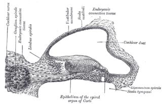
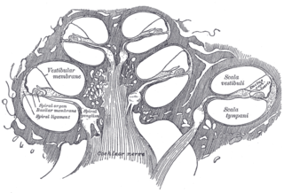
The spiral (cochlear) ganglion is a group of neuron cell bodies in the modiolus, the conical central axis of the cochlea. These bipolar neurons innervate the hair cells of the organ of Corti. They project their axons to the ventral and dorsal cochlear nuclei as the cochlear nerve, a branch of the vestibulocochlear nerve.
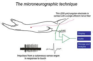
Microneurography is a neurophysiological method employed to visualize and record the traffic of nerve impulses that are conducted in peripheral nerves of waking human subjects. It can also be used in animal recordings. The method has been successfully employed to reveal functional properties of a number of neural systems, e.g. sensory systems related to touch, pain, and muscle sense as well as sympathetic activity controlling the constriction state of blood vessels. To study nerve impulses of an identified nerve, a fine tungsten needle microelectrode is inserted into the nerve and connected to a high input impedance differential amplifier. The exact position of the electrode tip within the nerve is then adjusted in minute steps until the electrode discriminates nerve impulses of interest. A unique feature and a significant strength of the microneurography method is that subjects are fully awake and able to cooperate in tests requiring mental attention, while impulses in a representative nerve fibre or set of nerve fibres are recorded, e.g. when cutaneous sense organs are stimulated or subjects perform voluntary precision movements.
The Hering–Breuer inflation reflex, named for Josef Breuer and Ewald Hering, is a reflex triggered to prevent the over-inflation of the lung. Pulmonary stretch receptors present on the wall of bronchi and bronchioles of the airways respond to excessive stretching of the lung during large inspirations.
Cutaneous innervation refers to an area of the skin which is supplied by a specific cutaneous nerve.

Group C nerve fibers are one of three classes of nerve fiber in the central nervous system (CNS) and peripheral nervous system (PNS). The C group fibers are unmyelinated and have a small diameter and low conduction velocity, whereas Groups A and B are myelinated. Group C fibers include postganglionic fibers in the autonomic nervous system (ANS), and nerve fibers at the dorsal roots. These fibers carry sensory information.
The Bezold–Jarisch reflex involves a variety of cardiovascular and neurological processes which cause hypopnea, hypotension and bradycardia in response to noxious stimuli detected in the cardiac ventricles. The reflex is named after Albert von Bezold and Adolf Jarisch Junior. The significance of the discovery is that it was the first recognition of a chemical (non-mechanical) reflex.
Group A nerve fibers are one of the three classes of nerve fiber as generally classified by Erlanger and Gasser. The other two classes are the group B nerve fibers, and the group C nerve fibers. Group A are heavily myelinated, group B are moderately myelinated, and group C are unmyelinated.

The Golgi tendon organ (GTO) is a proprioceptor – a type of sensory receptor that senses changes in muscle tension. It lies at the interface between a muscle and its tendon known as the musculotendinous junction also known as the myotendinous junction. It provides the sensory component of the Golgi tendon reflex.
References
- ↑ Guyton, Arthur C.; Hall, John Edward (2006). Textbook of Medical Physiology. Elsevier Saunders. ISBN 978-0-7216-0240-0.
- ↑ "Classification of Nerve Fibers". pharmacy180.com. Retrieved 2023-09-06.
| | This neuroanatomy article is a stub. You can help Wikipedia by expanding it. |