HARP (algorithm)

Last updated
HARP
Developer(s) Image Analysis and Communications Laboratory
Operating system Linux, Mac OS X, Windows
Type Cardiac Motion Tracking
Website HARP Overview (Software Download)

Harmonic phase (HARP) algorithm [1] is a medical image analysis technique capable of extracting and processing motion information from tagged magnetic resonance image (MRI) sequences. It was initially developed by N. F. Osman and J. L. Prince at the Image Analysis and Communications Laboratory at Johns Hopkins University. The method uses spectral peaks in the Fourier domain of tagged MRI, calculating the phase images of their inverse Fourier transforms, which are called harmonic phase (HARP) images. The motion of material points through time is then tracked, under the assumption that the HARP value of a fixed material point is time-invariant. The method is fast and accurate, and has been accepted as one of the most popular tagged MRI analysis methods in medical image processing.

Contents

Background

In cardiac magnetic resonance imaging, tagging techniques [2] [3] [4] [5] make it possible to capture and store the motion information of myocardium in vivo. MR tagging uses a special pulse sequence to create temporary features – tags in the myocardium. Tags deform together with the myocardium as the heart beats and are captured by MR imaging. Analysis of the motion of the tag features in many images taken from different orientations and at different times can be used to track material points in the myocardium. [6] [7] Tagged MRI is widely used to develop and refine models of normal and abnormal myocardial motion [8] [9] [10] [11] to better understand the correlation of coronary artery disease with myocardial motion abnormalities and the effects of treatment after myocardial infarction. However, suffered from long imaging and post-processing times, [12] tagged MRI was slow in entering into routine clinical use until the HARP algorithm was developed and published in 1999. [13]

Description

HARP processing

Demonstration of the HARP processing of a tagged cardiac MRI slice. (a) An MR image with vertical SPAMM tags. (b) shows the magnitude of its Fourier transform. By extracting the spectral peak inside the circle, a complex image is produced with a magnitude (c) and a phase (d). HARP Algorithm Processing.png
Demonstration of the HARP processing of a tagged cardiac MRI slice. (a) An MR image with vertical SPAMM tags. (b) shows the magnitude of its Fourier transform. By extracting the spectral peak inside the circle, a complex image is produced with a magnitude (c) and a phase (d).

A tagged MRI showing motion of a human heart is shown in the image (a). The effect of tagging can be described as a multiplication of the underlying image by a sinusoid tag pattern having a certain fundamental frequency, causing an amplitude modulation of the underlying image and replicating its Fourier transform into the pattern shown in (b).

HARP processing uses a bandpass filter to isolate one of the spectral peaks. For example, the circle drawn in (b) is the -3 dB isocontour of the bandpass filter used to process this data. Selection of the filters for optimal performance is discussed in this paper. [14] The inverse Fourier transform of the filtered image yields a complex harmonic image at image coordinates and time :

where is called the harmonic magnitude image and is called the harmonic phase image. The harmonic magnitude image in (c) extracted from a using the filter in (b) shows the geometry of the heart. And the harmonic phase image in (d) contains the motion of the myocardium in horizontal direction. In practice, tagged images from two directions (both horizontal and vertical, i.e., is 1 and 2) are processed to provide a 2D motion map in the image plane. Notice that the harmonic phase images are computed by taking the inverse tangent of the imaginary part divided by the real part of , such that the range of this computation is only in . In other words, d is only the wrapped value of the actual phase. We denote this principle value by ; it is mathematically related to the true phase by:

Either or might be called a harmonic phase (HARP) image, but only can be directly calculated and visualized. It is the basis for HARP tracking.

HARP tracking

Result of HARP tracking of a tagged cardiac MRI slice HARP Tracking.png
Result of HARP tracking of a tagged cardiac MRI slice

For a fixed material point with a HARP value, only one of the points sharing the same HARP value in a later time frame is the correct match. If the apparent motion is small from one image to the next, it is likely that the nearest of these points is the correct point. The tracking result is very accurate in this case.[ citation needed ]

Consider a material point located at at time . If is the apparent position of this point at time , we have:

The Newton–Raphson interactive method is used to find a solution, which is:

In practice, since is not available, is used in its place. This equation can be rewritten after a few derivations considering the "wrapping" relation between and .

The result of HARP tracking of one frame of cardiac MRI is shown in the figure. It is obtained by calculating both motions from horizontal direction and vertical direction, resulting in a 2D vector field showing the motion of every material point on the myocardium at this time frame.

The entire HARP algorithm takes only a few minutes to perform on a normal computer and the motion tracking result is accurate (with a typical error range of pixel). As a result, it is now widely adopted by the medical image analysis community as a standard processing technique for tagged MRI.

See also

Related Research Articles

<span class="mw-page-title-main">Spherical harmonics</span> Special mathematical functions defined on the surface of a sphere

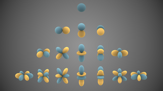
In mathematics and physical science, spherical harmonics are special functions defined on the surface of a sphere. They are often employed in solving partial differential equations in many scientific fields. The table of spherical harmonics contains a list of common spherical harmonics.

<span class="mw-page-title-main">Astronomical seeing</span> Atmospheric distortions of light

In astronomy, seeing is the degradation of the image of an astronomical object due to turbulence in the atmosphere of Earth that may become visible as blurring, twinkling or variable distortion. The origin of this effect is rapidly changing variations of the optical refractive index along the light path from the object to the detector. Seeing is a major limitation to the angular resolution in astronomical observations with telescopes that would otherwise be limited through diffraction by the size of the telescope aperture. Today, many large scientific ground-based optical telescopes include adaptive optics to overcome seeing.

<span class="mw-page-title-main">Canonical quantization</span> Process of converting a classical physical theory into one compatible with quantum mechanics

In physics, canonical quantization is a procedure for quantizing a classical theory, while attempting to preserve the formal structure, such as symmetries, of the classical theory to the greatest extent possible.

Phase-contrast imaging is a method of imaging that has a range of different applications. It measures differences in the refractive index of different materials to differentiate between structures under analysis. In conventional light microscopy, phase contrast can be employed to distinguish between structures of similar transparency, and to examine crystals on the basis of their double refraction. This has uses in biological, medical and geological science. In X-ray tomography, the same physical principles can be used to increase image contrast by highlighting small details of differing refractive index within structures that are otherwise uniform. In transmission electron microscopy (TEM), phase contrast enables very high resolution (HR) imaging, making it possible to distinguish features a few Angstrom apart.

<span class="mw-page-title-main">Hofstadter's butterfly</span> Fractal describing the theorised behaviour of electrons in a magnetic field

In condensed matter physics, Hofstadter's butterfly is a graph of the spectral properties of non-interacting two-dimensional electrons in a perpendicular magnetic field in a lattice. The fractal, self-similar nature of the spectrum was discovered in the 1976 Ph.D. work of Douglas Hofstadter and is one of the early examples of modern scientific data visualization. The name reflects the fact that, as Hofstadter wrote, "the large gaps [in the graph] form a very striking pattern somewhat resembling a butterfly."

<span class="mw-page-title-main">Magnetic resonance angiography</span> Group of techniques based on magnetic resonance imaging (MRI) to image blood vessels.

Magnetic resonance angiography (MRA) is a group of techniques based on magnetic resonance imaging (MRI) to image blood vessels. Magnetic resonance angiography is used to generate images of arteries in order to evaluate them for stenosis, occlusions, aneurysms or other abnormalities. MRA is often used to evaluate the arteries of the neck and brain, the thoracic and abdominal aorta, the renal arteries, and the legs.

In quantum mechanics, the energies of cyclotron orbits of charged particles in a uniform magnetic field are quantized to discrete values, called Landau levels. These levels are degenerate, with the number of electrons per level directly proportional to the strength of the applied magnetic field. It is named after the Soviet physicist Lev Landau.

In special functions, a topic in mathematics, spin-weighted spherical harmonics are generalizations of the standard spherical harmonics and—like the usual spherical harmonics—are functions on the sphere. Unlike ordinary spherical harmonics, the spin-weighted harmonics are U(1) gauge fields rather than scalar fields: mathematically, they take values in a complex line bundle. The spin-weighted harmonics are organized by degree l, just like ordinary spherical harmonics, but have an additional spin weights that reflects the additional U(1) symmetry. A special basis of harmonics can be derived from the Laplace spherical harmonics Ylm, and are typically denoted by sYlm, where l and m are the usual parameters familiar from the standard Laplace spherical harmonics. In this special basis, the spin-weighted spherical harmonics appear as actual functions, because the choice of a polar axis fixes the U(1) gauge ambiguity. The spin-weighted spherical harmonics can be obtained from the standard spherical harmonics by application of spin raising and lowering operators. In particular, the spin-weighted spherical harmonics of spin weight s = 0 are simply the standard spherical harmonics:

<span class="mw-page-title-main">Cardiac magnetic resonance imaging</span>

Cardiac magnetic resonance imaging, also known as cardiovascular MRI, is a magnetic resonance imaging (MRI) technology used for non-invasive assessment of the function and structure of the cardiovascular system. Conditions in which it is performed include congenital heart disease, cardiomyopathies and valvular heart disease, diseases of the aorta such as dissection, aneurysm and coarctation, coronary heart disease. It can also be used to look at pulmonary veins.

In mathematics, vector spherical harmonics (VSH) are an extension of the scalar spherical harmonics for use with vector fields. The components of the VSH are complex-valued functions expressed in the spherical coordinate basis vectors.

<span class="mw-page-title-main">Susceptibility weighted imaging</span>

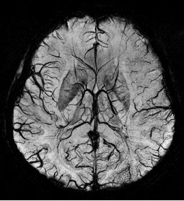
Susceptibility weighted imaging (SWI), originally called BOLD venographic imaging, is an MRI sequence that is exquisitely sensitive to venous blood, hemorrhage and iron storage. SWI uses a fully flow compensated, long echo, gradient recalled echo (GRE) pulse sequence to acquire images. This method exploits the susceptibility differences between tissues and uses the phase image to detect these differences. The magnitude and phase data are combined to produce an enhanced contrast magnitude image. The imaging of venous blood with SWI is a blood-oxygen-level dependent (BOLD) technique which is why it was referred to as BOLD venography. Due to its sensitivity to venous blood SWI is commonly used in traumatic brain injuries (TBI) and for high resolution brain venographies but has many other clinical applications. SWI is offered as a clinical package by Philips and Siemens but can be run on any manufacturer's machine at field strengths of 1.0 T, 1.5 T, 3.0 T and higher.

In the mathematical field of integral geometry, the Funk transform is an integral transform defined by integrating a function on great circles of the sphere. It was introduced by Paul Funk in 1911, based on the work of Minkowski (1904). It is closely related to the Radon transform. The original motivation for studying the Funk transform was to describe Zoll metrics on the sphere.

<span class="mw-page-title-main">Real-time MRI</span> Type of MRI

Real-time magnetic resonance imaging (RT-MRI) refers to the continuous monitoring ("filming") of moving objects in real time. Because MRI is based on time-consuming scanning of k-space, real-time MRI was possible only with low image quality or low temporal resolution. Using an iterative reconstruction algorithm these limitations have recently been removed: a new method for real-time MRI achieves a temporal resolution of 20 to 30 milliseconds for images with an in-plane resolution of 1.5 to 2.0 mm. Real-time MRI promises to add important information about diseases of the joints and the heart. In many cases MRI examinations may become easier and more comfortable for patients.

<span class="mw-page-title-main">Cardiac magnetic resonance imaging perfusion</span>

Cardiac magnetic resonance imaging perfusion, also known as stress CMR perfusion, is a clinical magnetic resonance imaging test performed on patients with known or suspected coronary artery disease to determine if there are perfusion defects in the myocardium of the left ventricle that are caused by narrowing of one or more of the coronary arteries.

<span class="mw-page-title-main">Philip Batchelor</span> British mathematician

Philip Batchelor, was a Swiss-British academic in the fields of mathematics and medical imaging.

In statistics, kernel Fisher discriminant analysis (KFD), also known as generalized discriminant analysis and kernel discriminant analysis, is a kernelized version of linear discriminant analysis (LDA). It is named after Ronald Fisher.

In pure and applied mathematics, quantum mechanics and computer graphics, a tensor operator generalizes the notion of operators which are scalars and vectors. A special class of these are spherical tensor operators which apply the notion of the spherical basis and spherical harmonics. The spherical basis closely relates to the description of angular momentum in quantum mechanics and spherical harmonic functions. The coordinate-free generalization of a tensor operator is known as a representation operator.

The assessment of the regional function of the heart is a good tool for early detection of deterioration in certain parts of the heart wall before a cardiac arrest is diagnosed. One of the most accurate measures of changes in regional function is the use of strain as a measure of the regional function of cardiac muscle.

<span class="mw-page-title-main">Fluctuation X-ray scattering</span>

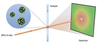
Fluctuation X-ray scattering (FXS) is an X-ray scattering technique similar to small-angle X-ray scattering (SAXS), but is performed using X-ray exposures below sample rotational diffusion times. This technique, ideally performed with an ultra-bright X-ray light source, such as a free electron laser, results in data containing significantly more information as compared to traditional scattering methods.

<span class="mw-page-title-main">Phase contrast magnetic resonance imaging</span>

Phase contrast magnetic resonance imaging (PC-MRI) is a specific type of magnetic resonance imaging used primarily to determine flow velocities. PC-MRI can be considered a method of Magnetic Resonance Velocimetry. It also provides a method of magnetic resonance angiography. Since modern PC-MRI is typically time-resolved, it provides a means of 4D imaging.

References

  1. Osman, N.F.; McVeigh, E.R.; Prince, J.L. (2000). "Imaging Heart Motion Using Harmonic Phase MRI". IEEE Trans Med Imaging . 19 (3): 186–202. CiteSeerX   10.1.1.649.7174 . doi:10.1109/42.845177. PMID   10875703. S2CID   6351307.
  2. Zerhouni, E.A.; Parish, D.M.; Rogers, W.J.; Yang, A.; Shapiro, E.P. (1988). "Human heart: tagging with MR imaging—a method for noninvasive assessment of myocardial motion". Radiology . 169 (1): 59–63. doi:10.1148/radiology.169.1.3420283. PMID   3420283.
  3. Axel, L.; Dougherty, L. (1989). "MR imaging of motion with spatial modulation of magnetization". Radiology . 171 (3): 841–845. doi:10.1148/radiology.171.3.2717762. PMID   2717762. S2CID   34168537.
  4. McVeigh, E.R.; Atalar, E. (1992). "Cardiac tagging with breath-hold cine MRI". Magn Reson Med . 28 (2): 318–327. doi:10.1002/mrm.1910280214. PMC   2041925 . PMID   1461130.
  5. Fischer, S.E.; McKinnon, G.C.; Maier, S.E.; Boesiger, P. (1993). "Improved myocardial tagging contrast". Magn Reson Med . 30 (2): 191–200. doi:10.1002/mrm.1910300207. PMID   8366800. S2CID   45146949.
  6. McVeigh, E.R. (1998). "Regional myocardial function". Cardiol Clin . 16 (2): 189–206. doi: 10.1016/s0733-8651(05)70008-4 . PMID   9627756.
  7. McVeigh, E.R. (1996). "MRI of myocardial function: motion tracking techniques". Magn Reson Imag . 14 (2): 137–150. doi: 10.1016/0730-725x(95)02009-i . PMID   8847969.
  8. Young, A.A.; Axel, L. (1992). "Three-dimensional motion and deformation of the heart wall: estimation with spatial modulation of magnetization—a model-based approach". Radiology . 185 (2): 241–247. doi:10.1148/radiology.185.1.1523316. PMID   1523316.
  9. Moore, C.; O'Dell, W.; McVeigh, E.R.; Zerhouni, E. (1992). "Calculation of three-dimensional left ventricular strains from biplanar tagged MR images". Magn Reson Imag . 2 (2): 165–175. doi:10.1002/jmri.1880020209. PMC   2041907 . PMID   1562767.
  10. Clark, N.R.; Reichek, N.; Bergey, P.; Hoffman, E.A.; Brownson, D.; Palmon, L.; Axel, L. (1991). "Circumferential myocardial shortening in the normal human left ventricle". Circulation . 84 (1): 67–74. doi: 10.1161/01.cir.84.1.67 . PMID   2060124.
  11. McVeigh, E.R.; Zerhouni, E.A. (1991). "Noninvasive measurements of transmural gradients in myocardial strain with MR imaging". Radiology . 180 (3): 677–683. doi:10.1148/radiology.180.3.1871278. PMC   2475677 . PMID   1871278.
  12. Budinger, T.F.; Berson, A.; McVeigh, E.R.; Pettigrew, R.I.; Pohost, G.M.; Watson, J.T.; Wickline, S.A. (1998). "Cardiac MR imaging: report of a working group sponsored by the National Heart, Lung, and Blood Institute". Radiology . 208 (3): 573–576. doi:10.1148/radiology.208.3.9722831. PMID   9722831.
  13. Osman, N.F.; Kerwin, W.S.; McVeigh, E.R.; Prince, J.L. (1999). "Cardiac Motion Tracking Using CINE Harmonic Phase (HARP) Magnetic Resonance Imaging". Magn Reson Med . 42 (6): 1048–1060. doi:10.1002/(sici)1522-2594(199912)42:6<1048::aid-mrm9>3.3.co;2-d. PMC   2570035 . PMID   10571926.
  14. Osman, N.F.; Prince, J.L. (1998). "Motion estimation from tagged MR images using angle images". Proc Int Conf Imag Proc : 704–708.