Related Research Articles

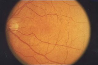
The retina is the innermost, light-sensitive layer of tissue of the eye of most vertebrates and some molluscs. The optics of the eye create a focused two-dimensional image of the visual world on the retina, which translates that image into electrical neural impulses to the brain to create visual perception. The retina serves a function analogous to that of the film or image sensor in a camera.

The thymus is a specialized primary lymphoid organ of the immune system. Within the thymus, Thymus cell lymphocytes or T cells mature. T cells are critical to the adaptive immune system, where the body adapts specifically to foreign invaders. The thymus is located in the upper front part of the chest, in the anterior superior mediastinum, behind the sternum, and in front of the heart. It is made up of two lobes, each consisting of a central medulla and an outer cortex, surrounded by a capsule.
Retinopathy is any damage to the retina of the eyes, which may cause vision impairment. Retinopathy often refers to retinal vascular disease, or damage to the retina caused by abnormal blood flow. Age-related macular degeneration is technically included under the umbrella term retinopathy but is often discussed as a separate entity. Retinopathy, or retinal vascular disease, can be broadly categorized into proliferative and non-proliferative types. Frequently, retinopathy is an ocular manifestation of systemic disease as seen in diabetes or hypertension. Diabetes is the most common cause of retinopathy in the U.S. as of 2008. Diabetic retinopathy is the leading cause of blindness in working-aged people. It accounts for about 5% of blindness worldwide and is designated a priority eye disease by the World Health Organization.

A cataract is an opacification of the lens of the eye which leads to a decrease in vision. Cataracts often develop slowly and can affect one or both eyes. Symptoms may include faded colors, blurry or double vision, halos around light, trouble with bright lights, and trouble seeing at night. This may result in trouble driving, reading, or recognizing faces. Poor vision caused by cataracts may also result in an increased risk of falling and depression. Cataracts cause half of all cases of blindness and 33% of visual impairment worldwide.

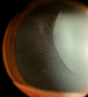
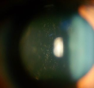
Floaters or eye floaters are sometimes visible deposits within the eye's vitreous humour, which is normally transparent, or between the vitreous and retina. Each floater can be measured by its size, shape, consistency, refractive index, and motility. They are also called muscae volitantes, or mouches volantes. The vitreous usually starts out transparent, but imperfections may gradually develop as one ages. The common type of floater, present in most people's eyes, is due to these degenerative changes of the vitreous. The perception of floaters, which may be annoying or problematic to some people, is known as myodesopsia, or, less commonly, as myodaeopsia, myiodeopsia, or myiodesopsia. It is not often treated, except in severe cases, where vitrectomy (surgery), laser vitreolysis, and medication may be effective.

The lens is a transparent biconvex structure in the eye that, along with the cornea, helps to refract light to be focused on the retina. By changing shape, it functions to change the focal length of the eye so that it can focus on objects at various distances, thus allowing a sharp real image of the object of interest to be formed on the retina. This adjustment of the lens is known as accommodation. Accommodation is similar to the focusing of a photographic camera via movement of its lenses. The lens is more flat on its anterior side than on its posterior side.
The vitreous body is the clear gel that fills the space between the lens and the retina of the eyeball of humans and other vertebrates. It is often referred to as the vitreous humor or simply "the vitreous".
The aqueous humour is a transparent watery fluid similar to plasma, but containing low protein concentrations. It is secreted from the ciliary body, a structure supporting the lens. It fills both the anterior and the posterior chambers of the eye, and is not to be confused with the vitreous humour, which is located in the space between the lens and the retina, also known as the posterior cavity or vitreous chamber.

Retinal detachment is a disorder of the eye in which the retina separates from the layer underneath. Symptoms include an increase in the number of floaters, flashes of light, and worsening of the outer part of the visual field. This may be described as a curtain over part of the field of vision. In about 7% of cases both eyes are affected. Without treatment permanent loss of vision may occur.

Fluorescein angiography (FA), fluorescent angiography (FAG), or fundus fluorescein angiography (FFA) is a technique for examining the circulation of the retina and choroid using a fluorescent dye and a specialized camera. Sodium fluorescein is added into the systemic circulation, the retina is illuminated with blue light at a wavelength of 490 nanometers, and an angiogram is obtained by photographing the fluorescent green light that is emitted by the dye. The fluorescein is administered intravenously in intravenous fluorescein angiography (IVFA) and orally in oral fluorescein angiography (OFA). The test is a dye tracing method.

Neurulation refers to the folding process in vertebrate embryos, which includes the transformation of the neural plate into the neural tube. The embryo at this stage is termed the neurula.

The ciliary body is a part of the eye that includes the ciliary muscle, which controls the shape of the lens, and the ciliary epithelium, which produces the aqueous humor. The aqueous humor is produced in the non-pigmented portion of the ciliary body. The ciliary body is part of the uvea, the layer of tissue that delivers oxygen and nutrients to the eye tissues. The ciliary body joins the ora serrata of the choroid to the root of the iris.

The human eye is a paired sense organ that reacts to light and allows vision. Rod and cone cells in the retina are photoreceptive cells which are able to detect visible light and convey this information to the brain. Eyes signal information which is used by the brain to elicit the perception of color, shape, depth, movement, and other features. The eye is part of the sensory nervous system.

Accommodation is the process by which the vertebrate eye changes optical power to maintain a clear image or focus on an object as its distance varies. In this, distances vary for individuals from the far point—the maximum distance from the eye for which a clear image of an object can be seen, to the near point—the minimum distance for a clear image. Accommodation usually acts like a reflex, including part of the accommodation-vergence reflex, but it can also be consciously controlled. Mammals, birds and reptiles vary their eyes' optical power by changing the form of the elastic lens using the ciliary body. Fish and amphibians vary the power by changing the distance between a rigid lens and the retina with muscles.
Aphakia is the absence of the lens of the eye, due to surgical removal, such as in cataract surgery, a perforating wound or ulcer, or congenital anomaly. It causes a loss of accommodation, high degree of farsightedness (hyperopia), and a deep anterior chamber. Complications include detachment of the vitreous or retina, and glaucoma.
Ectopia lentis is a displacement or malposition of the eye's crystalline lens from its normal location. A partial dislocation of a lens is termed lens subluxation or subluxated lens; a complete dislocation of a lens is termed lens luxation or luxated lens.
Intermediate uveitis is a form of uveitis localized to the vitreous and peripheral retina. Primary sites of inflammation include the vitreous of which other such entities as pars planitis, posterior cyclitis, and hyalitis are encompassed. Intermediate uveitis may either be an isolated eye disease or associated with the development of a systemic disease such as multiple sclerosis or sarcoidosis. As such, intermediate uveitis may be the first expression of a systemic condition. Infectious causes of intermediate uveitis include Epstein-Barr virus infection, Lyme disease, HTLV-1 virus infection, cat scratch disease, and hepatitis C.
David Anthony Newsome M.D. FARVO was a scientist, ophthalmologist, inventor, and author. He studied the treatment of age-related macular degeneration and proposed the usefulness of zinc supplements to slow the rate of vision loss from age-related macular degeneration.
Proliferative vitreoretinopathy (PVR) is a disease that develops as a complication of rhegmatogenous retinal detachment. PVR occurs in about 8–10% of patients undergoing primary retinal detachment surgery and prevents the successful surgical repair of rhegmatogenous retinal detachment. PVR can be treated with surgery to reattach the detached retina but the visual outcome of the surgery is very poor.
Sickle cell retinopathy is a major ocular complication of the sickle cell disease (SCD) which causes permanent loss of vision. Retinopathy can occur in sickling hemoglobinopathies like sickle cell disease, sickle cell C disease, and sickle cell thalassaemia disease.
References
- ↑ Sommer, F; Brandl, F; Weiser, B; Tesmar, J; Blunk, T; Göpferich, A (May 2009). "FACS as useful tool to study distinct hyalocyte populations". Exp Eye Res. 88 (5): 995–9. doi:10.1016/j.exer.2008.11.026. PMID 19073178.
- 1 2 Paulsen, Douglas F. (2010). "Chapter 24. Sense Organs". Histology and cell biology : examination and board review (5th ed.). Stamford, Conn.: Appleton & Lange. ISBN 978-0071476652.
- 1 2 3 Cunningham, Emmett T.; Paul Riordan-Eva (2011). "Chapter 1. Anatomy & Embryology of the Eye". Vaughan & Asbury's general ophthalmology (18th ed.). New York: McGraw-Hill Medical. ISBN 978-0071634205.
| This article about the eye is a stub. You can help Wikipedia by expanding it. |