
The tongue is a muscular organ in the mouth of a typical tetrapod. It manipulates food for mastication and swallowing as part of the digestive process, and is the primary organ of taste. The tongue's upper surface (dorsum) is covered by taste buds housed in numerous lingual papillae. It is sensitive and kept moist by saliva and is richly supplied with nerves and blood vessels. The tongue also serves as a natural means of cleaning the teeth. A major function of the tongue is the enabling of speech in humans and vocalization in other animals.

The facial nerve, also known as the seventh cranial nerve, cranial nerve VII, or simply CN VII, is a cranial nerve that emerges from the pons of the brainstem, controls the muscles of facial expression, and functions in the conveyance of taste sensations from the anterior two-thirds of the tongue. The nerve typically travels from the pons through the facial canal in the temporal bone and exits the skull at the stylomastoid foramen. It arises from the brainstem from an area posterior to the cranial nerve VI and anterior to cranial nerve VIII.

The glossopharyngeal nerve, also known as the ninth cranial nerve, cranial nerve IX, or simply CN IX, is a cranial nerve that exits the brainstem from the sides of the upper medulla, just anterior to the vagus nerve. Being a mixed nerve (sensorimotor), it carries afferent sensory and efferent motor information. The motor division of the glossopharyngeal nerve is derived from the basal plate of the embryonic medulla oblongata, whereas the sensory division originates from the cranial neural crest.

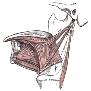
The hyoid bone is a horseshoe-shaped bone situated in the anterior midline of the neck between the chin and the thyroid cartilage. At rest, it lies between the base of the mandible and the third cervical vertebra.

The temporal bones are situated at the sides and base of the skull, and lateral to the temporal lobes of the cerebral cortex.

The suprahyoid muscles are four muscles located above the hyoid bone in the neck. They are the digastric, stylohyoid, geniohyoid, and mylohyoid muscles. They are all pharyngeal muscles, with the exception of the geniohyoid muscle. The digastric is uniquely named for its two bellies. Its posterior belly rises from the mastoid process of the cranium and slopes downward and forward. The anterior belly arises from the digastric fossa on the inner surface of the mandibular body, which slopes downward and backward. The two bellies connect at the intermediate tendon. The intermediate tendon passes through a connective tissue loop attached to the hyoid bone. The mylohyoid muscles are thin, flat muscles that form a sling inferior to the tongue supporting the floor of the mouth. The geniohyoids are short, narrow muscles that contact each other in the midline. The stylohyoids are long, thin muscles that are nearly parallel with the posterior belly of the digastric muscle.

The omohyoid muscle is a muscle that depresses the hyoid. It is located in the front of the neck, and consists of two bellies separated by an intermediate tendon. The omohyoid muscle is proximally attached to the scapula and distally attached to the hyoid bone, stabilising it. Its superior belly serves as the most lateral member of the infrahyoid muscles, located lateral to both the sternothyroid muscles and the thyrohyoid muscles.

The geniohyoid muscle is a narrow muscle situated superior to the medial border of the mylohyoid muscle. It is named for its passage from the chin to the hyoid bone.

The mylohyoid muscle or diaphragma oris is a paired muscle of the neck. It runs from the mandible to the hyoid bone, forming the floor of the oral cavity of the mouth. It is named after its two attachments near the molar teeth. It forms the floor of the submental triangle. It elevates the hyoid bone and the tongue, important during swallowing and speaking.

The stylohyoid muscle is a slender muscle, lying anterior and superior of the posterior belly of the digastric muscle. It is one of the suprahyoid muscles. It shares this muscle's innervation by the facial nerve, and functions to draw the hyoid bone backwards and elevate the tongue. Its origin is the styloid process of the temporal bone. It inserts on the body of the hyoid.

The hyoglossus, thin and quadrilateral, arises from the side of the body and from the whole length of the greater cornu of the hyoid bone, and passes almost vertically upward to enter the side of the tongue, between the styloglossus and the inferior longitudinal muscle of the tongue. It forms a part of the floor of submandibular triangle.
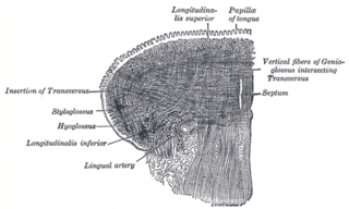
The inferior longitudinal muscle of tongue is an intrinsic muscle of the tongue. It is situated on the under surface of the tongue between the genioglossus and hyoglossus. It helps to move the tongue.

The middle pharyngeal constrictor is a fan-shaped muscle located in the neck. It is one of three pharyngeal constrictors. Similarly to the superior and inferior pharyngeal constrictor muscles, the middle pharyngeal constrictor is innervated by a branch of the vagus nerve through the pharyngeal plexus. The middle pharyngeal constrictor is smaller than the inferior pharyngeal constrictor muscle.
The chondroglossus muscle is a muscle of the tongue. It arises from the medial side of the lesser horn of the hyoid bone, before blending with intrinsic muscles of the tongue. It is supplied by the hypoglossal nerve.

The thyrohyoid membrane is a broad, fibro-elastic sheet of the larynx. It connects the upper border of the thyroid cartilage to the hyoid bone.

The lingual artery arises from the external carotid artery between the superior thyroid artery and facial artery. It can be located easily in the tongue.

The lingual septum consists of a vertical layer of fibrous tissue, extending throughout the entire length of the median plane of the tongue, though not quite reaching the dorsum. The lingual septum is closely associated with the hyoglossus membrane, allowing the binding of the tongue to the hyoid muscles.

The irregular bones are bones which, from their peculiar form, cannot be grouped as long, short, flat or sesamoid bones. Irregular bones serve various purposes in the body, such as protection of nervous tissue, affording multiple anchor points for skeletal muscle attachment, and maintaining pharynx and trachea support, and tongue attachment. They consist of cancellous tissue enclosed within a thin layer of compact bone. Irregular bones can also be used for joining all parts of the spinal column together. The spine is the place in the human body where the most irregular bones can be found. There are, in all, 33 irregular bones found here.

This article describes the anatomy of the head and neck of the human body, including the brain, bones, muscles, blood vessels, nerves, glands, nose, mouth, teeth, tongue, and throat.

The hyoid apparatus is the collective term used in veterinary anatomy for the bones which suspend the tongue and larynx. It consists of pairs of stylohyoid, thyrohyoid, epihyoid and ceratohyoid bones, and a single basihyoid bone. The hyoid apparatus resembles the shape of a trapeze, or a bent letter "H". The basihyoid bone lies within the muscle at the base of the tongue.