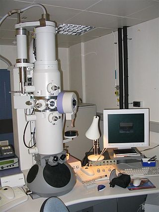
An electron microscope is a microscope that uses a beam of electrons as a source of illumination. They use electron optics that are analogous to the glass lenses of an optical light microscope to control the electron beam, for instance focusing them to produce magnified images or electron diffraction patterns. As the wavelength of an electron can be up to 100,000 times smaller than that of visible light, electron microscopes have a much higher resolution of about 0.1 nm, which compares to about 200 nm for light microscopes. Electron microscope may refer to:

Microscopy is the technical field of using microscopes to view objects and areas of objects that cannot be seen with the naked eye. There are three well-known branches of microscopy: optical, electron, and scanning probe microscopy, along with the emerging field of X-ray microscopy.

A scanning electron microscope (SEM) is a type of electron microscope that produces images of a sample by scanning the surface with a focused beam of electrons. The electrons interact with atoms in the sample, producing various signals that contain information about the surface topography and composition of the sample. The electron beam is scanned in a raster scan pattern, and the position of the beam is combined with the intensity of the detected signal to produce an image. In the most common SEM mode, secondary electrons emitted by atoms excited by the electron beam are detected using a secondary electron detector. The number of secondary electrons that can be detected, and thus the signal intensity, depends, among other things, on specimen topography. Some SEMs can achieve resolutions better than 1 nanometer.

Transmission electron microscopy (TEM) is a microscopy technique in which a beam of electrons is transmitted through a specimen to form an image. The specimen is most often an ultrathin section less than 100 nm thick or a suspension on a grid. An image is formed from the interaction of the electrons with the sample as the beam is transmitted through the specimen. The image is then magnified and focused onto an imaging device, such as a fluorescent screen, a layer of photographic film, or a detector such as a scintillator attached to a charge-coupled device or a direct electron detector.
Walter Cox McCrone Jr. was an American chemist who worked extensively on applications of polarized light microscopy and is sometimes characterized as the "father of modern microscopy". He was also an expert in electron microscopy, crystallography, ultra-microanalysis, and particle identification. In 1960 he founded the McCrone Research Institute, a non-profit educational and research organization for microscopy based in Chicago.

Confocal microscopy, most frequently confocal laser scanning microscopy (CLSM) or laser scanning confocal microscopy (LSCM), is an optical imaging technique for increasing optical resolution and contrast of a micrograph by means of using a spatial pinhole to block out-of-focus light in image formation. Capturing multiple two-dimensional images at different depths in a sample enables the reconstruction of three-dimensional structures within an object. This technique is used extensively in the scientific and industrial communities and typical applications are in life sciences, semiconductor inspection and materials science.

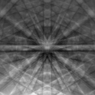
Electron backscatter diffraction (EBSD) is a scanning electron microscopy (SEM) technique used to study the crystallographic structure of materials. EBSD is carried out in a scanning electron microscope equipped with an EBSD detector comprising at least a phosphorescent screen, a compact lens and a low-light camera. In the microscope an incident beam of electrons hits a tilted sample. As backscattered electrons leave the sample, they interact with the atoms and are both elastically diffracted and lose energy, leaving the sample at various scattering angles before reaching the phosphor screen forming Kikuchi patterns (EBSPs). The EBSD spatial resolution depends on many factors, including the nature of the material under study and the sample preparation. They can be indexed to provide information about the material's grain structure, grain orientation, and phase at the micro-scale. EBSD is used for impurities and defect studies, plastic deformation, and statistical analysis for average misorientation, grain size, and crystallographic texture. EBSD can also be combined with energy-dispersive X-ray spectroscopy (EDS), cathodoluminescence (CL), and wavelength-dispersive X-ray spectroscopy (WDS) for advanced phase identification and materials discovery.

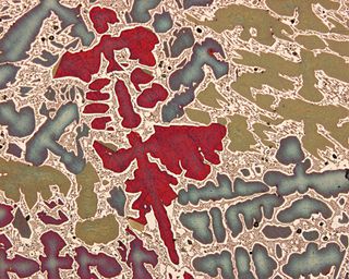
Metallography is the study of the physical structure and components of metals, by using microscopy.

George David William Smith FRS, FIMMM, FInstP, FRSC, CEng is a materials scientist with special interest in the study of the microstructure, composition and properties of engineering materials at the atomic level. He invented, together with Alfred Cerezo and Terry Godfrey, the Atom-Probe Tomograph in 1988.

Characterization, when used in materials science, refers to the broad and general process by which a material's structure and properties are probed and measured. It is a fundamental process in the field of materials science, without which no scientific understanding of engineering materials could be ascertained. The scope of the term often differs; some definitions limit the term's use to techniques which study the microscopic structure and properties of materials, while others use the term to refer to any materials analysis process including macroscopic techniques such as mechanical testing, thermal analysis and density calculation. The scale of the structures observed in materials characterization ranges from angstroms, such as in the imaging of individual atoms and chemical bonds, up to centimeters, such as in the imaging of coarse grain structures in metals.
Neuromorphology is the study of nervous system form, shape, and structure. The study involves looking at a particular part of the nervous system from a molecular and cellular level and connecting it to a physiological and anatomical point of view. The field also explores the communications and interactions within and between each specialized section of the nervous system. Morphology is distinct from morphogenesis. Morphology is the study of the shape and structure of biological organisms, while morphogenesis is the study of the biological development of the shape and structure of organisms. Therefore, neuromorphology focuses on the specifics of the structure of the nervous system and not the process by which the structure was developed. Neuromorphology and morphogenesis, while two different entities, are nonetheless closely linked.
Johan Sebastiaan Ploem is a Dutch microscopist and digital artist. He made significant contribution to the field of fluorescence microscopy, and invented reflection interference contrast microscopy.

The Royal Microscopical Society (RMS) is a learned society for the promotion of microscopy. It was founded in 1839 as the Microscopical Society of London making it the oldest organisation of its kind in the world. In 1866, the Society gained its royal charter and took its current name. Founded as a society of amateurs, its membership consists of individuals of all skill levels in numerous related fields from throughout the world. Every year since 1841, the Society has published its own scientific journal, the Journal of Microscopy, which contains peer-reviewed papers and book reviews. The Society is a registered charity that is dedicated to advancing science, developing careers and supporting wider understanding of science and microscopy through its Outreach activities.
Stereology is the three-dimensional interpretation of two-dimensional cross sections of materials or tissues. It provides practical techniques for extracting quantitative information about a three-dimensional material from measurements made on two-dimensional planar sections of the material. Stereology is a method that utilizes random, systematic sampling to provide unbiased and quantitative data. It is an important and efficient tool in many applications of microscopy. Stereology is a developing science with many important innovations being developed mainly in Europe. New innovations such as the proportionator continue to make important improvements in the efficiency of stereological procedures.

The Raman microscope is a laser-based microscopic device used to perform Raman spectroscopy. The term MOLE is used to refer to the Raman-based microprobe. The technique used is named after C. V. Raman, who discovered the scattering properties in liquids.
David Bernard Williams was the dean of the College of Engineering at the Ohio State University from 2011-2021. He was previously the fifth president of the University of Alabama in Huntsville in Huntsville, Alabama from March 2007 until April 2011, and Vice Provost for Research and Harold Chambers Senior Professor of Materials Science and Engineering at Lehigh University in Bethlehem, Pennsylvania.

A conservation scientist is a museum professional who works in the field of conservation science and whose focus is on the research of cultural heritage through scientific inquiry. Conservation scientists conduct applied scientific research and techniques to determine the material, chemical, and technical aspects of cultural heritage. The technical information conservation scientists gather is then used by conservator and curators to decide the most suitable conservation treatments for the examined object and/or adds to our knowledge about the object by providing answers about the material composition, fabrication, authenticity, and previous restoration treatments.
The American Microscopical Society (AMS) is a society of biologists dedicated to promoting the use of microscopy.

Image Analysis & Stereology (IAS) formerly Acta Stereologica, is a triannual peer-reviewed scientific journal published by an independent not-for-profit publisher DSKAS. It is the official journal of the International Society for Stereology & Image Analysis. The journal publishes articles of all fields of image analysis and processing.
The International Federation of Societies for Microscopy is an international non-governmental organization representing microscopy. It currently has 37 national members and 9 associate members, which are split into three regional committees, the Committee for Asia-Pacific Societies of Microscopy, the European Microscopy Society and the Interamerica Committee for Societies for EM.


