Related Research Articles

An epileptic seizure, informally known as a seizure, is a period of symptoms due to abnormally excessive or synchronous neuronal activity in the brain. Outward effects vary from uncontrolled shaking movements involving much of the body with loss of consciousness, to shaking movements involving only part of the body with variable levels of consciousness, to a subtle momentary loss of awareness. These episodes usually last less than two minutes and it takes some time to return to normal. Loss of bladder control may occur.
Clinical neurophysiology is a medical specialty that studies the central and peripheral nervous systems through the recording of bioelectrical activity, whether spontaneous or stimulated. It encompasses both research regarding the pathophysiology along with clinical methods used to diagnose diseases involving both central and peripheral nervous systems. Examinations in the clinical neurophysiology field are not limited to tests conducted in a laboratory. It is thought of as an extension of a neurologic consultation. Tests that are conducted are concerned with measuring the electrical functions of the brain, spinal cord, and nerves in the limbs and muscles. It can give the precise definition of site, the type and degree of the lesion, along with revealing the abnormalities that are in question. Due to these abilities, clinical neurophysiology is used to mainly help diagnose diseases rather than treat them.

Neurofeedback is a form of biofeedback that uses electrical potentials in the brain to reinforce desired brain states through operant conditioning. This process is non-invasive and typically collects brain activity data using electroencephalography (EEG). Several neurofeedback protocols exist, with potential additional benefit from use of quantitative electroencephalography (QEEG) or functional magnetic resonance imaging (fMRI) to localize and personalize treatment. Related technologies include functional near-infrared spectroscopy-mediated (fNIRS) neurofeedback, hemoencephalography biofeedback (HEG), and fMRI biofeedback.

An event-related potential (ERP) is the measured brain response that is the direct result of a specific sensory, cognitive, or motor event. More formally, it is any stereotyped electrophysiological response to a stimulus. The study of the brain in this way provides a noninvasive means of evaluating brain functioning.

A brain–computer interface (BCI), sometimes called a brain–machine interface (BMI) or smartbrain, is a direct communication pathway between the brain's electrical activity and an external device, most commonly a computer or robotic limb. BCIs are often directed at researching, mapping, assisting, augmenting, or repairing human cognitive or sensory-motor functions. They are often conceptualized as a human–machine interface that skips the intermediary component of the physical movement of body parts, although they also raise the possibility of the erasure of the discreteness of brain and machine. Implementations of BCIs range from non-invasive and partially invasive to invasive, based on how close electrodes get to brain tissue.
Neurotechnology encompasses any method or electronic device which interfaces with the nervous system to monitor or modulate neural activity.
Intraoperative neurophysiological monitoring (IONM) or intraoperative neuromonitoring is the use of electrophysiological methods such as electroencephalography (EEG), electromyography (EMG), and evoked potentials to monitor the functional integrity of certain neural structures during surgery. The purpose of IONM is to reduce the risk to the patient of iatrogenic damage to the nervous system, and/or to provide functional guidance to the surgeon and anesthesiologist.
Long-term or "continuous" video-electroencephalography (EEG) monitoring is a diagnostic technique commonly used in patients with epilepsy. It involves the long-term hospitalization of the patient, typically for days or weeks, during which brain waves are recorded via EEG and physical actions are continuously monitored by video. In epileptic patients, this technique is typically used to capture brain activity during seizures. The information gathered can be used for initial prognosis or long-term care management.
In neuroscience, single-unit recordings provide a method of measuring the electro-physiological responses of a single neuron using a microelectrode system. When a neuron generates an action potential, the signal propagates down the neuron as a current which flows in and out of the cell through excitable membrane regions in the soma and axon. A microelectrode is inserted into the brain, where it can record the rate of change in voltage with respect to time. These microelectrodes must be fine-tipped, impedance matching; they are primarily glass micro-pipettes, metal microelectrodes made of platinum, tungsten, iridium or even iridium oxide. Microelectrodes can be carefully placed close to the cell membrane, allowing the ability to record extracellularly.
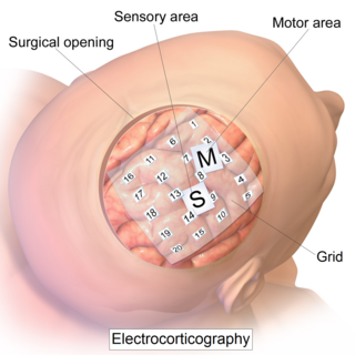
Electrocorticography (ECoG), a type of intracranial electroencephalography (iEEG), is a type of electrophysiological monitoring that uses electrodes placed directly on the exposed surface of the brain to record electrical activity from the cerebral cortex. In contrast, conventional electroencephalography (EEG) electrodes monitor this activity from outside the skull. ECoG may be performed either in the operating room during surgery or outside of surgery. Because a craniotomy is required to implant the electrode grid, ECoG is an invasive procedure.
Stereoelectroencephalography (SEEG) is the practice of recording electroencephalographic signals via depth electrodes. It may be used in patients with epilepsy not responding to medical treatment, and who are potential candidates to receive brain surgery in order to control seizures.
EEG-fMRI is a multimodal neuroimaging technique whereby EEG and fMRI data are recorded synchronously for the study of electrical brain activity in correlation with haemodynamic changes in brain during the electrical activity, be it normal function or associated with disorders.

The 10–20 system or International 10–20 system is an internationally recognized method to describe and apply the location of scalp electrodes in the context of an EEG exam, polysomnograph sleep study, or voluntary lab research. This method was developed to maintain standardized testing methods ensuring that a subject's study outcomes could be compiled, reproduced, and effectively analyzed and compared using the scientific method. The system is based on the relationship between the location of an electrode and the underlying area of the brain, specifically the cerebral cortex.
Electroencephalography (EEG) is a method to record an electrogram of the spontaneous electrical activity of the brain. The biosignals detected by EEG have been shown to represent the postsynaptic potentials of pyramidal neurons in the neocortex and allocortex. It is typically non-invasive, with the EEG electrodes placed along the scalp using the International 10–20 system, or variations of it. Electrocorticography, involving surgical placement of electrodes, is sometimes called "intracranial EEG". Clinical interpretation of EEG recordings is most often performed by visual inspection of the tracing or quantitative EEG analysis.

The neurotrophic electrode is an intracortical device designed to read the electrical signals that the brain uses to process information. It consists of a small, hollow glass cone attached to several electrically conductive gold wires. The term neurotrophic means "relating to the nutrition and maintenance of nerve tissue" and the device gets its name from the fact that it is coated with Matrigel and nerve growth factor to encourage the expansion of neurites through its tip. It was invented by neurologist Dr. Philip Kennedy and was successfully implanted for the first time in a human patient in 1996 by neurosurgeon Roy Bakay.
Cortical stimulation mapping (CSM) is a type of electrocorticography that involves a physically invasive procedure and aims to localize the function of specific brain regions through direct electrical stimulation of the cerebral cortex. It remains one of the earliest methods of analyzing the brain and has allowed researchers to study the relationship between cortical structure and systemic function. Cortical stimulation mapping is used for a number of clinical and therapeutic applications, and remains the preferred method for the pre-surgical mapping of the motor cortex and language areas to prevent unnecessary functional damage. There are also some clinical applications for cortical stimulation mapping, such as the treatment of epilepsy.

Joseph A. Sgro is an American mathematician, neurologist / neurophysiologist, and an engineering technologist / entrepreneur in the field of frame grabbers, high-speed cameras, smart cameras, image processors, computer vision, and machine vision and learning technologies.
A cortical implant is a subset of neuroprosthetics that is in direct connection with the cerebral cortex of the brain. By directly interfacing with different regions of the cortex, the cortical implant can provide stimulation to an immediate area and provide different benefits, depending on its design and placement. A typical cortical implant is an implantable microelectrode array, which is a small device through which a neural signal can be received or transmitted.

Fetal electroencephalography, also known as prenatal EEG includes any recording of electrical fluctuations arising from the brain of a fetus. Doctors and scientists use EEGs to detect and characterize brain activity, such as sleep states, potential seizures, or levels of a coma. EEG captures the electrical activity in the vicinity of the recording electrodes. The majority of the neural electrical activity arises from the flow of current from the cell bodies of pyramidal neurons to their apical dendrites, which become depolarized by excitatory inputs from other neurons. To record the most accurate signals, scientists try to minimize the distance between the recording electrode and the neural activity that they want to detect. Given the difficulty of attaching electrodes to a fetus inside a uterus, doctors and scientists use a variety of techniques to record fetal brain activity.
EEG analysis is exploiting mathematical signal analysis methods and computer technology to extract information from electroencephalography (EEG) signals. The targets of EEG analysis are to help researchers gain a better understanding of the brain; assist physicians in diagnosis and treatment choices; and to boost brain-computer interface (BCI) technology. There are many ways to roughly categorize EEG analysis methods. If a mathematical model is exploited to fit the sampled EEG signals, the method can be categorized as parametric, otherwise, it is a non-parametric method. Traditionally, most EEG analysis methods fall into four categories: time domain, frequency domain, time-frequency domain, and nonlinear methods. There are also later methods including deep neural networks (DNNs).
References
- ↑ A. Waziri; A. J. Claassen; R. M. Stuart; H. Arif; J. M. Schmidt; S. A. Mayer; N. Badjatia; L. L. Jull; E. S. Connolly; R. G. Emerson; L. J. Hirsch. (2009). Intracortical electroencephalography in acute brain injury. Annals of Neurology, Apr; 66(3):366–77.
- ↑ R. M. Stuart, A. Waziri, D. Weintraub, M. J. Schmidt, L. Fernandez, R. Helbok, P. Kurtz, K. Lee, N. Badjatia, E. S. C. R. Emersonand S. A. Mayer, L. J. Hirsch, and J. Claassen. (2010). Intracortical eeg for the detection of vasospasm in patients with poor-grade subarachnoid hemorrhage. Neurocrit Care, Jul 22.
{{cite book}}: CS1 maint: multiple names: authors list (link) - ↑ A. Waziri; H. Mehta; R. R. Goodman. (2008). Optimization of post-operative computerized tomographic imaging in patients with the implanted responsive neurostimulation system. Stereotactic Funct Neurosurg, May; 86(4):2037.
- ↑ A. Waziri; C. A. Schevon; J. Cappell; R. G. Emerson; M. GM; R. R. Goodman. (2009). Initial surgical experience with a dense cortical microarray in epileptic patients undergoing craniotomy for subdural electrode implantation. Neurosurgery, Mar; 64(3):540–45.
- ↑ R. Lewis; D. Shmueli; A. M. White. (2010). Deterministic Finite Automata in the Detection of Eeg Spikes and Seizures. The Joint Venture of the Ninth International Symposium on Intelligent Data Analysis (IDA), Tucson, Arizona, pages 103–113, May, 9–21.