Related Research Articles

Lichen planus (LP) is a chronic inflammatory and immune-mediated disease that affects the skin, nails, hair, and mucous membranes. It is not an actual lichen, and is only named that because it looks like one. It is characterized by polygonal, flat-topped, violaceous papules and plaques with overlying, reticulated, fine white scale, commonly affecting dorsal hands, flexural wrists and forearms, trunk, anterior lower legs and oral mucosa. Although there is a broad clinical range of LP manifestations, the skin and oral cavity remain as the major sites of involvement. The cause is unknown, but it is thought to be the result of an autoimmune process with an unknown initial trigger. There is no cure, but many different medications and procedures have been used in efforts to control the symptoms.
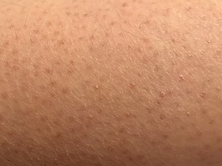
Keratosis pilaris (KP) is a common, autosomal dominant, genetic condition of the skin's hair follicles characterized by the appearance of possibly itchy, small, gooseflesh-like bumps, with varying degrees of reddening or inflammation. It most often appears on the outer sides of the upper arms, thighs, face, back, and buttocks; KP can also occur on the hands, and tops of legs, sides, or any body part except glabrous (hairless) skin. Often the lesions can appear on the face, which may be mistaken for acne.

A seborrheic keratosis is a non-cancerous (benign) skin tumour that originates from cells in the outer layer of the skin. Like liver spots, seborrheic keratoses are seen more often as people age.

Actinic keratosis (AK), sometimes called solar keratosis or senile keratosis, is a pre-cancerous area of thick, scaly, or crusty skin. Actinic keratosis is a disorder (-osis) of epidermal keratinocytes that is induced by ultraviolet (UV) light exposure (actin-). These growths are more common in fair-skinned people and those who are frequently in the sun. They are believed to form when skin gets damaged by UV radiation from the sun or indoor tanning beds, usually over the course of decades. Given their pre-cancerous nature, if left untreated, they may turn into a type of skin cancer called squamous cell carcinoma. Untreated lesions have up to a 20% risk of progression to squamous cell carcinoma, so treatment by a dermatologist is recommended.

Hyperkeratosis is thickening of the stratum corneum, often associated with the presence of an abnormal quantity of keratin, and also usually accompanied by an increase in the granular layer. As the corneum layer normally varies greatly in thickness in different sites, some experience is needed to assess minor degrees of hyperkeratosis.

Pityriasis rubra pilaris refers to a group of chronic disorders characterized by reddish orange, scaling plaques and keratotic follicular papules. Symptoms may include reddish-orange patches on the skin, severe flaking, uncomfortable itching, thickening of the skin on the feet and hands, and thickened bumps around hair follicles. For some, early symptoms may also include generalized swelling of the legs, feet and other parts of the body. PRP has a varied clinical progression and a varied rate of improvement. There is currently no known cause or cure for PRP.

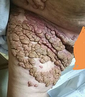
Keratoderma blennorrhagicum etymologically meaning keratinized (kerato-) skin (derma-) mucousy (blenno-) discharge (-rrhagia) are skin lesions commonly found on the palms and soles but which may spread to the scrotum, scalp and trunk. The lesions may resemble psoriasis.
Keratoderma is a hornlike skin condition.
Stomatitis nicotina is a diffuse white patch on the hard palate, usually caused by tobacco smoking, usually pipe or cigar smoking. It is painless, and it is caused by a response of the palatal oral mucosa to chronic heat. A more pronounced appearance can occur with reverse smoking, sometimes distinguished from stomatitis nicotina by the term reverse smoker's keratosis. While stomatitis nicotina that is caused by heat is not a premaligant condition, the condition that is caused by reverse smoking is premalignant.
Lichen spinulosus is a rare skin disorder characterized by follicular keratotic papules that are grouped into large patches. It is a variant of keratosis pilaris named for its resemblance to a patch of lichen.
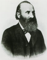
The Koebner phenomenon or Köbner phenomenon, also called the Koebner response or the isomorphic response, attributed to Heinrich Köbner, is the appearance of skin lesions on lines of trauma. The Koebner phenomenon may result from either a linear exposure or irritation. Conditions demonstrating linear lesions after a linear exposure to a causative agent include: molluscum contagiosum, warts and toxicodendron dermatitis. Warts and molluscum contagiosum lesions can be spread in linear patterns by self-scratching ("auto-inoculation"). Toxicodendron dermatitis lesions are often linear from brushing up against the plant. Causes of the Koebner phenomenon that are secondary to scratching rather than an infective or chemical cause include vitiligo, psoriasis, lichen planus, lichen nitidus, pityriasis rubra pilaris, and keratosis follicularis.

Hidrocystoma is an adenoma of the sweat glands.
Nevus lipomatosus (cutaneous) superficialis is characterized by soft, yellowish papules or cerebriform plaques, usually of the buttock or thigh, less often of the ear or scalp, with a wrinkled rather than warty surface. It is usually congenital in origin or appears within the first three decades.
Keratosis pilaris atropicans includes many forms of keratosis pilaris with cicatricial alopecia. Variants include keratosis pilaris atrophicans faciei, atrophoderma vermiculatum, keratosis follicularis spinulosa decalvans, and ichthyosis follicularis.
Keratosis follicularis spinulosa decalvans is a rare X-linked disorder described by Siemens in 1926, a disease that begins in infancy with keratosis pilaris localized on the face, then evolves to more diffuse involvement.
Keratosis punctata of the palmar creases is a common skin disorder that occurs most often in black patients, with skin lesions that are 1 to 5mm depressions filled with a comedo-like keratinous plug.
Ectodermal dysplasia with corkscrew hairs is a skin condition with salient features including exaggerated pili torti, scalp keloids, follicular plugging, keratosis pilaris, xerosis, eczema, palmoplantar keratoderma, syndactyly, onychodysplasia and conjunctival neovascularization.
A hydrocarbon keratosis is a precancerous keratotic skin lesion that occurs in people who have been occupationally exposed to polycyclic aromatic hydrocarbons.
A chronic scar keratosis is a precancerous skin lesion that arises within a long-standing scar.
A PUVA keratosis is a precancerous keratotic skin lesion that arises from exposure to psoralen plus ultraviolet A light therapy.
References
- 1 2 James, William; Berger, Timothy; Elston, Dirk (2005). Andrews' Diseases of the Skin: Clinical Dermatology. (10th ed.). Saunders. ISBN 0-7216-2921-0.
- ↑ Freedberg, et al. (2003). Fitzpatrick's Dermatology in General Medicine. (6th ed.). McGraw-Hill. ISBN 0-07-138076-0.
- ↑ Rapini, Ronald P.; Bolognia, Jean L.; Jorizzo, Joseph L. (2007). Dermatology: 2-Volume Set. St. Louis: Mosby. p. 522. ISBN 978-1-4160-2999-1.