Related Research Articles

Radiology is the medical specialty that uses medical imaging to diagnose diseases and guide treatment within the bodies of humans and other animals. It began with radiography, but today it includes all imaging modalities. This includes technologies that use no ionizing electromagnetic radiation, such as ultrasonography and magnetic resonance imaging), as well as others that do use radiation, such as computed tomography (CT), fluoroscopy, and nuclear medicine including positron emission tomography (PET). Interventional radiology is the performance of usually minimally invasive medical procedures with the guidance of imaging technologies such as those mentioned above.
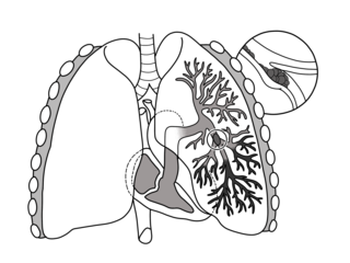
Pulmonary embolism (PE) is a blockage of an artery in the lungs by a substance that has moved from elsewhere in the body through the bloodstream (embolism). Symptoms of a PE may include shortness of breath, chest pain particularly upon breathing in, and coughing up blood. Symptoms of a blood clot in the leg may also be present, such as a red, warm, swollen, and painful leg. Signs of a PE include low blood oxygen levels, rapid breathing, rapid heart rate, and sometimes a mild fever. Severe cases can lead to passing out, abnormally low blood pressure, obstructive shock, and sudden death.
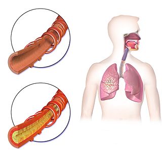
Hemoptysis or haemoptysis is the discharge of blood or blood-stained mucus through the mouth coming from the bronchi, larynx, trachea, or lungs. It does not necessarily involve coughing. In other words, it is the airway bleeding. This can occur with lung cancer, infections such as tuberculosis, bronchitis, or pneumonia, and certain cardiovascular conditions. Hemoptysis is considered massive at 300 mL. In such cases, there are always severe injuries. The primary danger comes from choking, rather than blood loss.
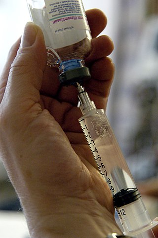
An air embolism, also known as a gas embolism, is a blood vessel blockage caused by one or more bubbles of air or other gas in the circulatory system. Air can be introduced into the circulation during surgical procedures, lung over-expansion injury, decompression, and a few other causes. In flora, air embolisms may also occur in the xylem of vascular plants, especially when suffering from water stress.

Angiography or arteriography is a medical imaging technique used to visualize the inside, or lumen, of blood vessels and organs of the body, with particular interest in the arteries, veins, and the heart chambers. Modern angiography is performed by injecting a radio-opaque contrast agent into the blood vessel and imaging using X-ray based techniques such as fluoroscopy. With time-of-flight (TOF) magnetic ressonance it is no longer necessary to use a contrast.
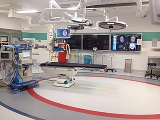
Interventional radiology (IR) is a medical specialty that performs various minimally-invasive procedures using medical imaging guidance, such as x-ray fluoroscopy, computed tomography, magnetic resonance imaging, or ultrasound. IR performs both diagnostic and therapeutic procedures through very small incisions or body orifices. Diagnostic IR procedures are those intended to help make a diagnosis or guide further medical treatment, and include image-guided biopsy of a tumor or injection of an imaging contrast agent into a hollow structure, such as a blood vessel or a duct. By contrast, therapeutic IR procedures provide direct treatment—they include catheter-based medicine delivery, medical device placement, and angioplasty of narrowed structures.

Pulmonary angiography is a medical fluoroscopic procedure used to visualize the pulmonary arteries and much less frequently, the pulmonary veins. It is a minimally invasive procedure performed most frequently by an interventional radiologist or interventional cardiologist to visualise the arteries of the lungs.

An inferior vena cava filter is a medical device made of metal that is implanted by vascular surgeons or interventional radiologists into the inferior vena cava to prevent a life-threatening pulmonary embolism (PE) or venous thromboembolism (VTE).

Uterine artery embolization is a procedure in which an interventional radiologist uses a catheter to deliver small particles that block the blood supply to the uterine body. The procedure is primarily done for the treatment of uterine fibroids and adenomyosis. Compared to surgical treatment for fibroids such as a hysterectomy, in which a woman's uterus is removed, uterine artery embolization may be beneficial in women who wish to retain their uterus. Other reasons for uterine artery embolization are postpartum hemorrhage and uterine arteriovenous malformations.
In chest radiography, the Westermark sign is a sign that represents a focus of oligemia (hypovolemia) seen distal to a pulmonary embolism (PE). While the chest x-ray is normal in the majority of PE cases, the Westermark sign is seen in 2% of patients.
George Getzel Cohen was a South African-Australian radiologist.

Computed tomography angiography is a computed tomography technique used for angiography—the visualization of arteries and veins—throughout the human body. Using contrast injected into the blood vessels, images are created to look for blockages, aneurysms, dissections, and stenosis. CTA can be used to visualize the vessels of the heart, the aorta and other large blood vessels, the lungs, the kidneys, the head and neck, and the arms and legs. CTA can also be used to localise arterial or venous bleed of the gastrointestinal system.

A CT pulmonary angiogram (CTPA) is a medical diagnostic test that employs computed tomography (CT) angiography to obtain an image of the pulmonary arteries. Its main use is to diagnose pulmonary embolism (PE). It is a preferred choice of imaging in the diagnosis of PE due to its minimally invasive nature for the patient, whose only requirement for the scan is an intravenous line.
Ground-glass opacity (GGO) is a finding seen on chest x-ray (radiograph) or computed tomography (CT) imaging of the lungs. It is typically defined as an area of hazy opacification (x-ray) or increased attenuation (CT) due to air displacement by fluid, airway collapse, fibrosis, or a neoplastic process. When a substance other than air fills an area of the lung it increases that area's density. On both x-ray and CT, this appears more grey or hazy as opposed to the normally dark-appearing lungs. Although it can sometimes be seen in normal lungs, common pathologic causes include infections, interstitial lung disease, and pulmonary edema.

Medical imaging in pregnancy may be indicated because of pregnancy complications, intercurrent diseases or routine prenatal care.

Aidoc Medical is an Israeli technology company that develops computer-aided simple triage and notification systems. Aidoc has obtained FDA and CE mark approval for its stroke, pulmonary embolism, cervical fracture, intracranial hemorrhage, intra-abdominal free gas, and incidental pulmonary embolism algorithms.
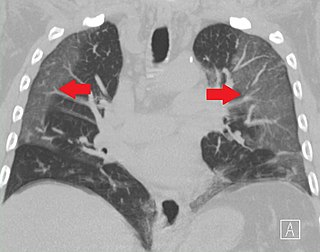
Fleischner sign is a radiological sign that aids the diagnosis of pulmonary embolism. The sign indicates the dilatation of the proximal pulmonary arteries due to pulmonary embolism. It was named after Felix Fleischner, who first described it. The Fleishner sign is seen both on X-ray and CT scan of chest/thorax.
Juxtaphrenic peak sign is a radiographic sign seen in lobar collapse or after lobectomy of the lung. This sign was first described by Katten and colleagues in 1980, and therefore, it is also called Katten's sign. The juxtaphrenic peak is most commonly caused due to the traction from the inferior accessory fissure. The prevalence of the juxtaphrenic peak sign increases gradually during the weeks after lobectomy of the lung.
Dr. Morris Simon, MB, BCH, (1926–2005) was a South African-born American radiologist, professor, and inventor. His medical practice was based primarily at Beth Israel Deaconess Medical Center, Boston, where he specialized in chest radiology. He is also credited with a number of medical inventions, including a flexible filter for dissolving blood clots, and innovations that streamlined patient care and records holding.
The black pleura sign is a radiological feature observed in pulmonary alveolar microlithiasis (PAM), a rare lung disorder characterized by the accumulation of tiny calcium phosphate deposits, known as microliths, within the alveoli. This sign appears as a thin, dark (lucent) line beneath the ribs on imaging studies, contrasting with the diffusely dense, calcified lung parenchyma.
References
- ↑ "Eurorad.org". Eurorad - Brought to you by the ESR. Retrieved 14 July 2021.
- 1 2 Kumaresh, Athiyappan; Kumar, Mitesh; Dev, Bhawna; Gorantla, Rajani; Sai, PM Venkata; Thanasekaraan, Vijayalakshmi (31 July 2015). "Back to Basics – 'Must Know' Classical Signs in Thoracic Radiology". Journal of Clinical Imaging Science. 5: 43. doi: 10.4103/2156-7514.161977 . ISSN 2156-7514. PMC 4541161 . PMID 26312141.
- ↑ Chiarenza, Alessandra; Esposto Ultimo, Luca; Falsaperla, Daniele; Travali, Mario; Foti, Pietro Valerio; Torrisi, Sebastiano Emanuele; Schisano, Matteo; Mauro, Letizia Antonella; Sambataro, Gianluca; Basile, Antonio; Vancheri, Carlo; Palmucci, Stefano (4 December 2019). "Chest imaging using signs, symbols, and naturalistic images: a practical guide for radiologists and non-radiologists". Insights into Imaging. 10 (1): 114. doi: 10.1186/s13244-019-0789-4 . ISSN 1869-4101. PMC 6893008 . PMID 31802270.