
Histology, also known as microscopic anatomy or microanatomy, is the branch of biology which studies the microscopic anatomy of biological tissues. Histology is the microscopic counterpart to gross anatomy, which looks at larger structures visible without a microscope. Although one may divide microscopic anatomy into organology, the study of organs, histology, the study of tissues, and cytology, the study of cells, modern usage places these topics under the field of histology. In medicine, histopathology is the branch of histology that includes the microscopic identification and study of diseased tissue. In the field of paleontology, the term paleohistology refers to the histology of fossil organisms.

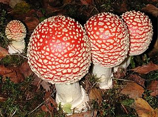
Ascomycota division or phylum of the kingdom Fungi that, together with the Basidiomycota, form the subkingdom Dikarya. Its members are commonly known as the sac fungi or ascomycetes. It is the largest phylum of Fungi, with over 64,000 species. The defining feature of this fungal group is the "ascus", a microscopic sexual structure in which nonmotile spores, called ascospores, are formed. However, some species of the Ascomycota are asexual, meaning that they do not have a sexual cycle and thus do not form asci or ascospores. Familiar examples of sac fungi include morels, truffles, brewer's yeast and baker's yeast, dead man's fingers, and cup fungi. The fungal symbionts in the majority of lichens such as Cladonia belong to the Ascomycota.
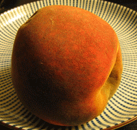
A mold or mould is a fungus that grows in the form of multicellular filaments called hyphae. In contrast, fungi that can adopt a single-celled growth habit are called yeasts.

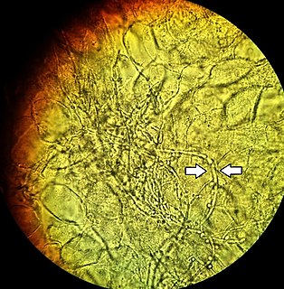
A hypha is a long, branching filamentous structure of a fungus, oomycete, or actinobacterium. In most fungi, hyphae are the main mode of vegetative growth, and are collectively called a mycelium.
A mycorrhiza is a symbiotic association between a fungus and a plant. The term mycorrhiza refers to the role of the fungus in the plant's rhizosphere, its root system. Mycorrhizae play important roles in plant nutrition, soil biology and soil chemistry.

In biology, cell theory is the historic scientific theory, now universally accepted, that living organisms are made up of cells, that they are the basic structural/organizational unit of all organisms, and that all cells come from pre-existing cells. Cells are the basic unit of structure in all organisms and also the basic unit of reproduction. With continual improvements made to microscopes over time, magnification technology advanced enough to discover cells in the 17th century. This discovery is largely attributed to Robert Hooke, and began the scientific study of cells, known as cell biology. Over a century later, many debates about cells began amongst scientists. Most of these debates involved the nature of cellular regeneration, and the idea of cells as a fundamental unit of life. Cell theory was eventually formulated in 1839. This is usually credited to Matthias Schleiden and Theodor Schwann. However, many other scientists like Rudolf Virchow contributed to the theory. It was an important step in the movement away from spontaneous generation.

Zygomycota, or zygote fungi, is a former division or phylum of the kingdom Fungi. The members are now part of two phyla the Mucoromycota and Zoopagomycota. Approximately 1060 species are known. They are mostly terrestrial in habitat, living in soil or on decaying plant or animal material. Some are parasites of plants, insects, and small animals, while others form symbiotic relationships with plants. Zygomycete hyphae may be coenocytic, forming septa only where gametes are formed or to wall off dead hyphae. Zygomycota is no longer recognised as it was not believed to be truly monophyletic
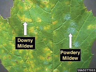
Mildew is a form of fungus. It is distinguished from its closely related counterpart, mold, largely by its color: moulds appear in shades of black, blue, red, and green, whereas mildew is white. It appears as a thin, superficial growth consisting of minute hyphae produced especially on living plants or organic matter such as wood, paper or leather. Both mould and mildew produce distinct offensive odors, and both have been identified as the cause of certain human ailments.

A microscope slide is a thin flat piece of glass, typically 75 by 26 mm and about 1 mm thick, used to hold objects for examination under a microscope. Typically the object is mounted (secured) on the slide, and then both are inserted together in the microscope for viewing. This arrangement allows several slide-mounted objects to be quickly inserted and removed from the microscope, labeled, transported, and stored in appropriate slide cases or folders etc.

Bromothymol blue is a pH indicator. It is mostly used in applications that require measuring substances that would have a relatively neutral pH. A common use is for measuring the presence of carbonic acid in a liquid. It is typically sold in solid form as the sodium salt of the acid indicator.

Histopathology refers to the microscopic examination of tissue in order to study the manifestations of disease. Specifically, in clinical medicine, histopathology refers to the examination of a biopsy or surgical specimen by a pathologist, after the specimen has been processed and histological sections have been placed onto glass slides. In contrast, cytopathology examines free cells or tissue micro-fragments.

In botany and mycology, a haustorium is a rootlike structure or a structure that grows into or around another structure to absorb water or nutrients. In botany, this may refer to a cotyledon, or to the root of a parasitic plant that penetrates the host's tissue and draws nutrients from it. In mycology, it refers to the appendage or portion of a parasitic fungus, which performs a similar function. Microscopic haustoria penetrate the host plant's cell wall and siphon nutrients from the space between the cell wall and plasma membrane but do not penetrate the membrane itself. Larger haustoria do this at the tissue level.
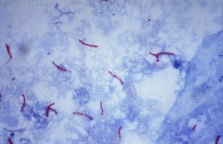
Ziehl-Neelsen staining is a type of Acid-fast stain, first introduced by Paul Ehrlich. Ziehl–Neelsen staining is a bacteriological stain used to identify acid-fast organisms, mainly Mycobacteria. It is named for two German doctors who modified the stain: the bacteriologist Franz Ziehl (1859–1926) and the pathologist Friedrich Neelsen (1854–1898).

Saprolegnia is a genus of water moulds often called cotton moulds because of the characteristic white or grey fibrous patches they form. Current taxonomy puts Saprolegnia as a genus of the heterokonts in the order Saprolegniales.
The KOH Test for Candida albicans, also known as a potassium hydroxide preparation or KOH prep, is a quick, inexpensive fungal test to differentiate dermatophytes and Candida albicans symptoms from other skin disorders like psoriasis and eczema.

A clamp connection is a hook-like structure formed by growing hyphal cells of certain fungi. It is created to ensure each cell, or segment of hypha separated by septa, receives a set of differing nuclei, which are obtained through mating of hyphae of differing sexual types. It is used to create genetic variation within the hypha much like the mechanisms found in crozier during sexual reproduction.
Microfungi are fungi— eukaryotic organisms such as molds, mildews and rusts— which exhibit tube tip-growth and have cell walls composed of chitin, a polymer of N-acetylglucosamine. Microfungi are an artificial, paraphyletic group, distinguished from macrofungi only by the absence of a large, multicellular fruiting body. They are ubiquitous in all terrestrial and freshwater and marine environments, and grow in plants, soil, water, insects, cattle rumens, hair, and skin. Most of the fungal body consists of microscopic threads, called hyphae, extending through the substrate in which it grows. The mycelia of microfungi produce spores that are carried by the air, spreading the fungus.
The Kinyoun method or Kinyoun stain, developed by Joseph J. Kinyoun, is a procedure used to stain acid-fast species of the bacterial genera Mycobacterium and Nocardia and the apicomplexan genus Cryptosporidium. It is a variation of a method developed by Robert Koch in 1882. Certain species of bacteria have a waxy lipid called mycolic acid, in their cell walls which allow them to be stained with Acid-Fast better than a Gram-Stain. The unique ability of mycobacteria to resist decolorization byacid-alcohol is why they are termed acid-fast. It involves the application of a primary stain, a decolorizer (acid-alcohol), and a counterstain. Unlike the Ziehl-Neelsen stain, the Kinyoun method of staining does not require heating. In the Ziehl-Neelsen stain, heat acts as a physical mordant while phenol acts as the chemical mordant. Since the Kinyoun stain is a cold method, the concentration of carbol fuschin used is increased.

Sporoidiobolus salmonicolor is a yeast-like fungus in the Basidiomycota more commonly known by the name of its asexual yeast-like state, Sporobolomyces salmonicolor. It is generally considered a Biosafety Risk Group 1 fungus; however isolates of S. salmonicolor have been recovered from cerebrospinal fluid, infected skin, a nasal polyp, lymphadenitis and a case of endophthalmitis. It has also been reported in AIDS-related infections. The fungus exists predominantly in the asexual state as a unicellular, haploid yeast yet this species can sometimes produce a sexual state when conjugation of compatible yeast cells occurs. The asexual form consists of a characteristic, pink, ballistosporic yeast. Ballistoconidia are borne from slender extensions of the cell known as sterigma, and are forcibly ejected into the air upon maturity. Levels of airborne yeast cells peak during the night and are abundant in areas of decaying leaves and grains. Three varieties of Sporobolomyces salmonicolor have been described; S. salmonicolor var. albus, S. salmonicolor var. fischerii, and S. salmonicolor var. salmoneus.

Dolipore septa are specialized dividing walls between cells (septa) found in almost all species of fungi in the phylum Basidiomycota. Unlike most fungal septa, they have a barrel-shaped swelling around their central pore, which is about 0.1–0.2 µm wide. This structure is typically capped at either end by specialized membranes, called "parenthesomes" or simply "pore caps".
