Nociception is the sensory nervous system's process of encoding noxious stimuli. In nociception, intense chemical, mechanical, or thermal stimulation of sensory nerve cells called nociceptors produces a signal that travels along a chain of nerve fibers via the spinal cord to the brain. Nociception triggers a variety of physiological and behavioral responses and usually results in a subjective experience, or perception, of pain in sentient beings.

In humans, vocal cords, also known as vocal folds or voice reeds are folds of tissue in the throat that are key in creating sounds through vocalization. The size of vocal cords affects the pitch of voice. Open when breathing and vibrating for speech or singing, the folds are controlled via the recurrent laryngeal branch of the vagus nerve. They are composed of twin infoldings of mucous membrane stretched horizontally, from back to front, across the larynx. They vibrate, modulating the flow of air being expelled from the lungs during phonation.

The retina is the innermost, light-sensitive layer of tissue of the eye of most vertebrates and some molluscs. The optics of the eye create a focused two-dimensional image of the visual world on the retina, which translates that image into electrical neural impulses to the brain to create visual perception. The retina serves a function analogous to that of the film or image sensor in a camera.

Neuropil is any area in the nervous system composed of mostly unmyelinated axons, dendrites and glial cell processes that forms a synaptically dense region containing a relatively low number of cell bodies. The most prevalent anatomical region of neuropil is the brain which, although not completely composed of neuropil, does have the largest and highest synaptically-concentrated areas of neuropil in the body. For example, the neocortex and olfactory bulb both contain neuropil.

A fibroblast is a type of biological cell that synthesizes the extracellular matrix and collagen, produces the structural framework (stroma) for animal tissues, and plays a critical role in wound healing. Fibroblasts are the most common cells of connective tissue in animals.

In biology, the extracellular matrix (ECM) is a three-dimensional network of extracellular macromolecules, such as collagen, enzymes, and glycoproteins, that provide structural and biochemical support to surrounding cells. Because multicellularity evolved independently in different multicellular lineages, the composition of ECM varies between multicellular structures; however, cell adhesion, cell-to-cell communication and differentiation are common functions of the ECM.

The brown algae, comprising the class Phaeophyceae, are a large group of multicellular algae, including many seaweeds located in colder waters within the Northern Hemisphere. Most brown algae live in marine environments, where they play an important role both as food and as a potential habitat. For instance, Macrocystis, a kelp of the order Laminariales, may reach 60 m (200 ft) in length and forms prominent underwater kelp forests. Kelp forests like these contain a high level of biodiversity. Another example is Sargassum, which creates unique floating mats of seaweed in the tropical waters of the Sargasso Sea that serve as the habitats for many species. Many brown algae, such as members of the order Fucales, commonly grow along rocky seashores. Some members of the class, such as kelps, are used by humans as food.

Santiago Ramón y Cajal was a Spanish neuroscientist and pathologist, specializing in neuroanatomy, particularly the histology of the central nervous system. He and Camillo Golgi received the Nobel Prize in Physiology or Medicine in 1906, with Ramón y Cajal thereby becoming the first person of Spanish origin to win a scientific Nobel Prize. His original investigations of the microscopic structure of the brain made him a pioneer of modern neuroscience. Hundreds of his drawings illustrating the delicate arborizations of brain cells are still in use for educational and training purposes.
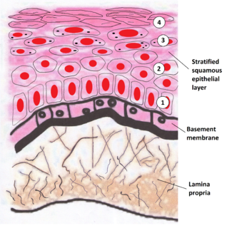
The lamina propria is a thin layer of connective tissue that forms part of the moist linings known as mucous membranes or mucosa, which line various tubes in the body, such as the respiratory tract, the gastrointestinal tract, and the urogenital tract.

The superior colliculus is a structure lying on the roof of the mammalian midbrain. In non-mammalian vertebrates the homologous structure, is known as the optic tectum or optic lobe. The adjective form tectal is commonly used for both structures.
The basement membrane is a thin, fibrous, extracellular matrix that separates the lining of an internal or external body surface from underlying connective tissue in metazoans (animals). This surface may be epithelium, mesothelium and endothelium
The oral mucosa is the mucous membrane lining the inside of the mouth. It comprises stratified squamous epithelium, termed "oral epithelium", and an underlying connective tissue termed lamina propria. The oral cavity has sometimes been described as a mirror that reflects the health of the individual. Changes indicative of disease are seen as alterations in the oral mucosa lining the mouth, which can reveal systemic conditions, such as diabetes or vitamin deficiency, or the local effects of chronic tobacco or alcohol use. The oral mucosa tends to heal faster and with less scar formation compared to the skin. The underlying mechanism remains unknown, but research suggests that extracellular vesicles might be involved.

Descemet's membrane is the basement membrane that lies between the corneal proper substance, also called stroma, and the endothelial layer of the cornea. It is composed of different kinds of collagen than the stroma. The endothelial layer is located at the posterior of the cornea. Descemet's membrane, as the basement membrane for the endothelial layer, is secreted by the single layer of squamous epithelial cells that compose the endothelial layer of the cornea.
Gut-associated lymphoid tissue (GALT) is a component of the mucosa-associated lymphoid tissue (MALT) which works in the immune system to protect the body from invasion in the gut.

Electrosurgery is the application of a high-frequency alternating polarity, electrical current to biological tissue as a means to cut, coagulate, desiccate, or fulgurate tissue.. Its benefits include the ability to make precise cuts with limited blood loss. Electrosurgical devices are frequently used during surgical operations helping to prevent blood loss in hospital operating rooms or in outpatient procedures.
The capillary lamina of choroid or choriocapillaris is a layer of capillaries that is immediately adjacent to Bruch's membrane in the choroid. The choriocapillaris was first described in man by Hovius in 1702, although it was not so named until 1838, by Eschricht. Passera (1896) described its form as star-shaped, radiating capillaries beneath the pigment epithelium of the retina, and Duke-Elder and Wybar (1961) have emphasized its nature as a network of capillaries in one plane.. The choriocapillaris serves multiple functions that include sustaining the photoreceptors, filtering waste produced in the outer retina and regulating the temperature of macula. The capillary wall is permeable to plasma proteins which is probably of great importance for the supply of vitamin A to the pigment epithelium.
Collagen VI (ColVI) is a type of collagen primarily associated with the extracellular matrix of skeletal muscle. ColVI maintains regularity in muscle function and stabilizes the cell membrane. It is synthesized by a complex, multistep pathway that leads to the formation of a unique network of linked microfilaments located in the extracellular matrix (ECM). ColVI plays a vital role in numerous cell types, including chondrocytes, neurons, myocytes, fibroblasts, and cardiomyocytes. ColVI molecules are made up of three alpha chains: α1(VI), α2(VI), and α3(VI). It is encoded by 6 genes: COL6A1, COL6A2, COL6A3, COL6A4, COL6A5, and COL6A6. The chain lengths of α1(VI) and α2(VI) are about 1,000 amino acids. The chain length of α3(VI) is roughly a third larger than those of α1(VI) and α2(VI), and it consists of several spliced variants within the range of 2,500 to 3,100 amino acids.
The lamina is the most peripheral neuropil of the insect visual system. There are twelve distinct neuron classes in the lamina: the lamina monopolar cells L1-L5, two GABAergic feedback neurons, two wide-field feedback neurons, lamina intrinsic amacrine neurons (Lai) and the T1 basket cell. The outer photoreceptors, R1-R6, terminate in the lamina, where they form tetrad synapses with L1, L2, L3, and Lai.

A central nervous system primitive neuroectodermal tumor, often abbreviated as PNET, supratentorial PNET, or CNS-PNET, is one of the 3 types of embryonal central nervous system tumors defined by the World Health Organization. It is considered an embryonal tumor because it arises from cells partially differentiated or still undifferentiated from birth. Those cells are usually neuroepithelial cells, stem cells destined to turn into glia or neurons. It can occur anywhere within the spinal cord and cerebrum and can have multiple sites of origins, with a high probability of metastasis through cerebrospinal fluid (CSF).