Infrared radiation (IR), sometimes called infrared light, is electromagnetic radiation (EMR) with longer wavelengths than those of visible light, and is therefore generally invisible to the human eye, although IR at wavelengths up to 1050 nanometers (nm)s from specially pulsed lasers can be seen by humans under certain conditions. IR wavelengths extend from the nominal red edge of the visible spectrum at 700 nanometers, to 1 millimeter (300 GHz). Most of the thermal radiation emitted by objects near room temperature is infrared. As with all EMR, IR carries radiant energy and behaves both like a wave and like its quantum particle, the photon.

Spectroscopy is the study of the interaction between matter and electromagnetic radiation. Historically, spectroscopy originated through the study of visible light dispersed according to its wavelength, by a prism. Later the concept was expanded greatly to include any interaction with radiative energy as a function of its wavelength or frequency, predominantly in the electromagnetic spectrum, though matter waves and acoustic waves can also be considered forms of radiative energy; recently, with tremendous difficulty, even gravitational waves have been associated with a spectral signature in the context of LIGO and laser interferometry. Spectroscopic data are often represented by an emission spectrum, a plot of the response of interest as a function of wavelength or frequency.

Raman spectroscopy ; named after Indian physicist Sir C. V. Raman) is a spectroscopic technique used to observe vibrational, rotational, and other low-frequency modes in a system. Raman spectroscopy is commonly used in chemistry to provide a structural fingerprint by which molecules can be identified.
Cavity ring-down spectroscopy (CRDS) is a highly sensitive optical spectroscopic technique that enables measurement of absolute optical extinction by samples that scatter and absorb light. It has been widely used to study gaseous samples which absorb light at specific wavelengths, and in turn to determine mole fractions down to the parts per trillion level. The technique is also known as cavity ring-down laser absorption spectroscopy (CRLAS).
Ellipsometry is an optical technique for investigating the dielectric properties of thin films. Ellipsometry measures the change of polarization upon reflection or transmission and compares it to a model.
Resonance Raman spectroscopy is a Raman spectroscopy technique in which the incident photon energy is close in energy to an electronic transition of a compound or material under examination. The frequency coincidence can lead to greatly enhanced intensity of the Raman scattering, which facilitates the study of chemical compounds present at low concentrations.
A staring array, staring-plane array, focal-plane array (FPA), or focal-plane is an image sensing device consisting of an array of light-sensing pixels at the focal plane of a lens. FPAs are used most commonly for imaging purposes, but can also be used for non-imaging purposes such as spectrometry, LIDAR, and wave-front sensing.
Far-infrared laser or terahertz laser is a laser with output wavelength in between 30-1000 µm, in the far infrared or terahertz frequency band of the electromagnetic spectrum.
Digital ICE or Digital Image Correction and Enhancement is a set of technologies related to producing an altered image in a variety of frequency spectra. The objective of these technologies is to render an image more usable by Fourier or other filtering techniques. These technologies were most actively advanced in the 1960s and early 1970s in the fields of strategic reconnaissance and medical electronics.
Photothermal microspectroscopy (PTMS), alternatively known as photothermal temperature fluctuation (PTTF), is derived from two parent instrumental techniques: infrared spectroscopy and atomic force microscopy (AFM). In one particular type of AFM, known as scanning thermal microscopy (SThM), the imaging probe is a sub-miniature temperature sensor, which may be a thermocouple or a resistance thermometer. This same type of detector is employed in a PTMS instrument, enabling it to provide AFM/SThM images: However, the chief additional use of PTMS is to yield infrared spectra from sample regions below a micrometer, as outlined below.

Laser ablation electrospray ionization (LAESI) is an ambient ionization method for mass spectrometry that combines laser ablation from a mid-infrared (mid-IR) laser with a secondary electrospray ionization (ESI) process. The mid-IR laser is used to generate gas phase particles which are then ionized through interactions with charged droplets from the ESI source. LAESI was developed by Professor Akos Vertes and Dr. Peter Nemes in 2007 and is now marketed commercially by Protea Biosciences, Inc. LAESI is a novel ionization source for mass spectrometry (MS) that has been used to perform MS imaging of plants, tissues, cell pellets, and even single cells. In addition, LAESI has been used to analyze historic documents and untreated biofluids such as urine and blood. The technique of LAESI is performed at atmospheric pressure and therefore overcomes many of the obstacles of traditional MS techniques, including extensive and invasive sample preparation steps and the use of high vacuum.
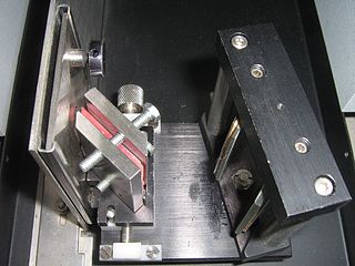
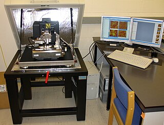
The technique of vibrational analysis with scanning probe microscopy allows probing vibrational properties of materials at the submicrometer scale, and even of individual molecules. This is accomplished by integrating scanning probe microscopy (SPM) and vibrational spectroscopy. This combination allows for much higher spatial resolution than can be achieved with conventional Raman/FTIR instrumentation. The technique is also nondestructive, requires non-extensive sample preparation, and provides more contrast such as intensity contrast, polarization contrast and wavelength contrast, as well as providing specific chemical information and topography images simultaneously.

Interband cascade lasers (ICLs) are a type of laser diode that can produce coherent radiation over a large part of the mid-infrared region of the electromagnetic spectrum. They are fabricated from epitaxially-grown semiconductor heterostructures composed of layers of indium arsenide (InAs), gallium antimonide (GaSb), aluminum antimonide (AlSb), and related alloys. These lasers are similar to quantum cascade lasers (QCLs) in several ways. Like QCLs, ICLs employ the concept of bandstructure engineering to achieve an optimized laser design and reuse injected electrons to emit multiple photons. However, in ICLs, photons are generated with interband transitions, rather than the intersubband transitions used in QCLs. Consequently, the rate at which the carriers injected into the upper laser subband thermally relax to the lower subband is determined by interband Auger, radiative, and Shockley-Read carrier recombination. These processes typically occur on a much slower time scale than the longitudinal optical phonon interactions that mediates the intersubband relaxation of injected electrons in mid-IR QCLs. The use of interband transitions allows laser action in ICLs to be achieved at lower electrical input powers than is possible with QCLs.

The NIRSpec is one of the four scientific instruments which will be flown on the James Webb Space Telescope (JWST). The JWST is the follow-on mission to the Hubble Space Telescope (HST) and is developed to receive more information about the origins of the universe by observing infrared light from the first stars and galaxies. In comparison to HST, its instruments will allow looking further back in time and will study the so-called Dark Ages during which the universe was opaque, about 150 to 800 million years after the Big Bang.
A. R. Forouhi and I. Bloomer deduced dispersion equations for the refractive index, n, and extinction coefficient, k, which were published in 1986 and 1988. The 1986 publication relates to amorphous materials, while the 1988 publication relates to crystalline. Subsequently, in 1991, their work was included as a chapter in “The Handbook of Optical Constants”. The Forouhi–Bloomer dispersion equations describe how photons of varying energies interact with thin films. When used with a spectroscopic reflectometry tool, the Forouhi–Bloomer dispersion equations specify n and k for amorphous and crystalline materials as a function of photon energy E. Values of n and k as a function of photon energy, E, are referred to as the spectra of n and k, which can also be expressed as functions of wavelength of light, λ, since E = hc/λ. The symbol h represents Planck’s constant and c, the speed of light in vacuum. Together, n and k are often referred to as the “optical constants” of a material.
AFM-IR is one of a family of techniques that are derived from a combination of two parent instrumental techniques; infrared spectroscopy and scanning probe microscopy (SPM). The term was first used to denote a method that combined a tuneable free electron laser with an atomic force microscope equipped with a sharp probe that measured the local absorption of infrared light by a sample; it required that the sample be coupled to an infrared-transparent prism and be less than 1μm thick. It improved the spatial resolution of photothermal AFM-based techniques from microns to circa 100 nm.

Nano-FTIR is a scanning probe technique that can be considered as a combination of two techniques: Fourier transform infrared spectroscopy (FTIR) and scattering-type scanning near-field optical microscopy (s-SNOM). As s-SNOM, nano-FTIR is based on atomic-force microscopy (AFM), where a sharp tip is illuminated by an external light source and the tip-scattered light is detected as a function of tip position. A typical nano-FTIR setup thus consists of an atomic force microscope, a broadband infrared light source used for tip illumination, and a Michelson interferometer acting as Fourier transform spectrometer. In nano-FTIR, the sample stage is placed in one of the interferometer arms, which allows for recording both amplitude and phase of the detected light. Scanning the tip allows for performing hyperspectral imaging with nanoscale spatial resolution determined by the tip apex size. The use of broadband infrared sources enables the acquisition of continuous spectra, which is a distinctive feature of nano-FTIR compared to s-SNOM. Nano-FTIR is capable of performing infrared (IR) spectroscopy of materials in ultrasmall quantities and with nanoscale spatial resolution. The detection of a single molecular complex and the sensitivity to a single monolayer has been shown. Recording infrared spectra as a function of position can be used for nanoscale mapping of the sample chemical composition, performing a local ultrafast IR spectroscopy and analyzing the nanoscale intermolecular coupling, among others. A spatial resolution of 10 nm to 20 nm is routinely achieved
Origins Space Telescope (OST) is a concept study for the Far-Infrared Surveyor space telescope mission. Still a preliminary concept in formulation, it will be presented to the United States Decadal Survey in 2019 for a possible selection to NASA's Flagship Program. The OST would provide an array of new tools for studying star formation and the energetics and physical state of the interstellar medium within the Milky Way using infrared radiation and new spectroscopic capabilities.