External links
- Patello-femoral Pain Syndrome {This article has a good image}
| Lateral retinaculum | |
|---|---|
| Identifiers | |
| TA98 | A03.6.08.017 |
| TA2 | 2624 |
| FMA | 44590 44590, 44590 |
| Anatomical terminology | |
The lateral retinaculum is the fibrous tissue on the lateral (outer) side of the kneecap (patella). The kneecap has both a medial (on the inner aspect) and a lateral (on the outer side) retinaculum, and these help to support the kneecap in its position in relation to the femur bone underneath it.
The lateral retinaculum is an extension of the fibrous 'aponeurosis' of the vastus lateralis muscle (itself a part of the quadriceps muscles making up the 'lap').

In humans and other primates, the knee joins the thigh with the leg and consists of two joints: one between the femur and tibia, and one between the femur and patella. It is the largest joint in the human body. The knee is a modified hinge joint, which permits flexion and extension as well as slight internal and external rotation. The knee is vulnerable to injury and to the development of osteoarthritis.

The median nerve is a nerve in humans and other animals in the upper limb. It is one of the five main nerves originating from the brachial plexus.
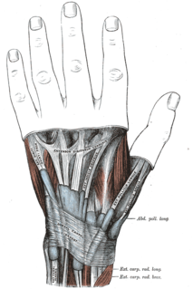
In human anatomy, extensor carpi radialis brevis is a muscle in the forearm that acts to extend and abduct the wrist. It is shorter and thicker than its namesake extensor carpi radialis longus which can be found above the proximal end of the extensor carpi radialis brevis.

In human anatomy, the peroneus longus is a superficial muscle in the lateral compartment of the leg, and acts to evert and plantarflex the ankle.

The patella, also known as the kneecap, is a flat, rounded triangular bone which articulates with the femur and covers and protects the anterior articular surface of the knee joint. The patella is found in many tetrapods, such as mice, cats, birds and dogs, but not in whales, or most reptiles.
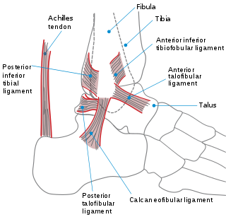
The ankle, or the talocrural region, is the region where the foot and the leg meet. The ankle includes three joints: the ankle joint proper or talocrural joint, the subtalar joint, and the inferior tibiofibular joint. The movements produced at this joint are dorsiflexion and plantarflexion of the foot. In common usage, the term ankle refers exclusively to the ankle region. In medical terminology, "ankle" can refer broadly to the region or specifically to the talocrural joint.
The extensor digiti minimi is a slender muscle of the forearm, placed on the ulnar side of the extensor digitorum communis, with which it is generally connected.

The tibial nerve is a branch of the sciatic nerve. The tibial nerve passes through the popliteal fossa to pass below the arch of soleus.

The extensor digitorum longus is a pennate muscle, situated at the lateral part of the front of the leg.

The extensor digitorum brevis muscle is a muscle on the upper surface of the foot that helps extend digits 2 through 4.
The peroneus brevismuscle lies under cover of the peroneus longus, and is the shorter and smaller of the peroneus muscles.

The tensor fasciae latae is a muscle of the thigh. Together with the gluteus maximus, it acts on the iliotibial band and is continuous with the iliotibial tract, which attaches to the tibia. The muscle assists in keeping the balance of the pelvis while standing, walking, or running.
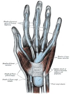
The flexor retinaculum is a fibrous band on the palmar side of the hand near the wrist. It arches over the carpal bones of the hands, covering them and forming the carpal tunnel.
The fascia lata is the deep fascia of the thigh. It encloses the thigh muscles and forms the outer limit of the fascial compartments of thigh, which are internally separated by intermuscular septa. The fascia lata is thickened at its lateral side where it forms the iliotibial tract, a structure that runs to the tibia and serves as a site of muscle attachment.

The peroneal retinacula are fibrous retaining bands which bind down the tendons of the peroneus longus and brevis as they run across the side of the ankle..
The flexor retinaculum of foot is a strong fibrous band in the foot.
The extensor retinaculum is an anatomical term for the thickened part of the antebrachial fascia that holds the tendons of the extensor muscles in place. It is located on the back of the forearm, just proximal to the hand. It is continuous with the palmar carpal ligament, which is located on the anterior side of the forearm.
The superior extensor retinaculum of the foot is the upper part of the extensor retinaculum of foot which extends from the ankle to the heelbone.

In the human body, the carpal tunnel or carpal canal is the passageway on the palmar side of the wrist that connects the forearm to the hand.
A lateral release is a surgical procedure to release tight capsular structures on the outer aspect of the kneecap (patella). This is usually performed because of knee pain related to the kneecap being pulled over to the outer (lateral) side and not being able to run properly in the centre of the groove of the femur bone as the knee bends and straightens. The procedure is also known as a 'lateral retinacular release'.