The human leg, in the general word sense, is the entire lower limb of the human body, including the foot, thigh or sometimes even the hip or gluteal region. However, the definition in human anatomy refers only to the section of the lower limb extending from the knee to the ankle, also known as the crus or, especially in non-technical use, the shank. Legs are used for standing, and all forms of locomotion including recreational such as dancing, and constitute a significant portion of a person's mass. Female legs generally have greater hip anteversion and tibiofemoral angles, but shorter femur and tibial lengths than those in males.

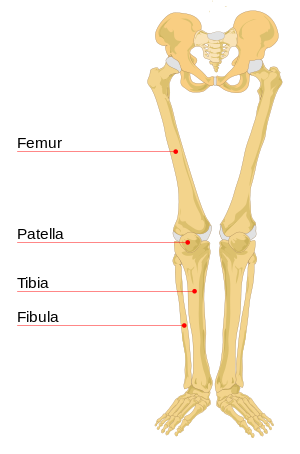
The femur, or thigh bone, is the proximal bone of the hindlimb in tetrapod vertebrates. The head of the femur articulates with the acetabulum in the pelvic bone forming the hip joint, while the distal part of the femur articulates with the tibia (shinbone) and patella (kneecap), forming the knee joint. By most measures the two femurs are the strongest bones of the body, and in humans, the largest and thickest.

In humans and other primates, the knee joins the thigh with the leg and consists of two joints: one between the femur and tibia, and one between the femur and patella. It is the largest joint in the human body. The knee is a modified hinge joint, which permits flexion and extension as well as slight internal and external rotation. The knee is vulnerable to injury and to the development of osteoarthritis.

The tibia, also known as the shinbone or shankbone, is the larger, stronger, and anterior (frontal) of the two bones in the leg below the knee in vertebrates ; it connects the knee with the ankle. The tibia is found on the medial side of the leg next to the fibula and closer to the median plane. The tibia is connected to the fibula by the interosseous membrane of leg, forming a type of fibrous joint called a syndesmosis with very little movement. The tibia is named for the flute tibia. It is the second largest bone in the human body, after the femur. The leg bones are the strongest long bones as they support the rest of the body.

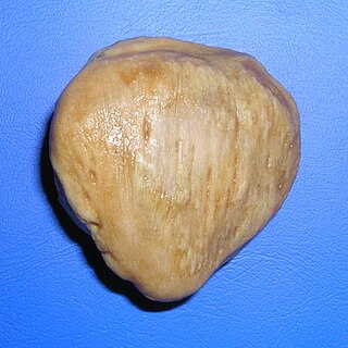
The patella, also known as the kneecap, is a flat, rounded triangular bone which articulates with the femur and covers and protects the anterior articular surface of the knee joint. The patella is found in many tetrapods, such as mice, cats, birds and dogs, but not in whales, or most reptiles.
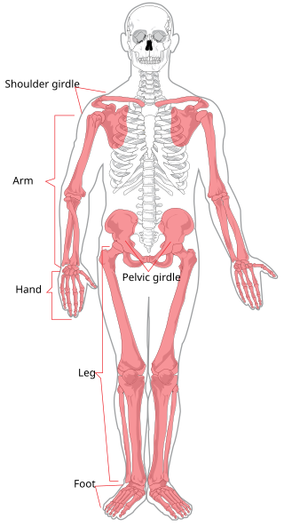
The appendicular skeleton is the portion of the skeleton of vertebrates consisting of the bones that support the appendages. There are 126 bones. The appendicular skeleton includes the skeletal elements within the limbs, as well as supporting shoulder girdle and pelvic girdle. The word appendicular is the adjective of the noun appendage, which itself means a part that is joined to something larger.
Attenuated patella alta is an extremely rare condition affecting mobility and leg strength. It is characterized by an unusually small knee cap (patella) that develops out of and above the joint. Typically, as the knee cap sits in the joint, it is stimulated to growth by abrasion from the opposing bones. When not situated properly in the joint, the knee cap does not experience such stimulation and remains small and undeveloped. Note that the cartilage under and around the kneecap is eight times smoother than ice, so "abrasion" may not be the best term.
The posterior cruciate ligament (PCL) is a ligament in each knee of humans and various other animals. It works as a counterpart to the anterior cruciate ligament (ACL). It connects the posterior intercondylar area of the tibia to the medial condyle of the femur. This configuration allows the PCL to resist forces pushing the tibia posteriorly relative to the femur.
The rectus femoris muscle is one of the four quadriceps muscles of the human body. The others are the vastus medialis, the vastus intermedius, and the vastus lateralis. All four parts of the quadriceps muscle attach to the patella by the quadriceps tendon.

The stifle joint is a complex joint in the hind limbs of quadruped mammals such as the sheep, horse or dog. It is the equivalent of the human knee and is often the largest synovial joint in the animal's body. The stifle joint joins three bones: the femur, patella, and tibia. The joint consists of three smaller ones: the femoropatellar joint, medial femorotibial joint, and lateral femorotibial joint.

Patellar tendon rupture is a tear of the tendon that connects the knee cap (patella) to the tibia. Often there is sudden onset of pain and walking is difficult. In a complete rupture, the ability to extend that knee is decreased. A pop may be felt when it occurs.

The patellar tendon is the distal portion of the common tendon of the quadriceps femoris, which is continued from the patella to the tibial tuberosity. It is also sometimes called the patellar ligament as it forms a bone to bone connection when the patella is fully ossified.
The tuberosity of the tibia or tibial tuberosity or tibial tubercle is an elevation on the proximal, anterior aspect of the tibia, just below where the anterior surfaces of the lateral and medial tibial condyles end.
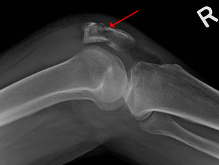
A patella fracture is a break of the kneecap. Symptoms include pain, swelling, and bruising to the front of the knee. A person may also be unable to walk. Complications may include injury to the tibia, femur, or knee ligaments.

The articular capsule of the knee joint is the wide and lax joint capsule of the knee. It is thin in front and at the side, and contains the patella, ligaments, menisci, and bursae of the knee. The capsule consists of an inner synovial membrane, and an outer fibrous membrane separated by fatty deposits anteriorly and posteriorly.
In human anatomy, the quadriceps tendon works with the quadriceps muscle to extend the leg. All four parts of the quadriceps muscle attach to the shin via the patella, where the quadriceps tendon becomes the patellar ligament. It attaches the quadriceps to the top of the patella, which in turn is connected to the shin from its bottom by the patellar ligament. A tendon connects muscle to bone, while a ligament connects bone to bone.

The deep fascia of leg, or crural fascia forms a complete investment to the muscles, and is fused with the periosteum over the subcutaneous surfaces of the bones.

A patellar dislocation is a knee injury in which the patella (kneecap) slips out of its normal position. Often the knee is partly bent, painful and swollen. The patella is also often felt and seen out of place. Complications may include a patella fracture or arthritis.

Good conformation in the limbs leads to improved movement and decreased likelihood of injuries. Large differences in bone structure and size can be found in horses used for different activities, but correct conformation remains relatively similar across the spectrum. Structural defects, as well as other problems such as injuries and infections, can cause lameness, or movement at an abnormal gait. Injuries to and problems with horse legs can be relatively minor, such as stocking up, which causes swelling without lameness, or quite serious. Even leg injuries that are not immediately fatal may still be life-threatening to horses, as their bodies are adapted to bear weight on all four legs and serious problems can result if this is not possible.
Bipartite patella is a condition where the patella, or kneecap, is composed of two separate bones. Instead of fusing together as normally occurs in early childhood, the bones of the patella remain separated. The condition occurs in approximately 1–2% of the population and is no more likely to occur in males than females. It is often asymptomatic and most commonly diagnosed as an incidental finding, with about 2% of cases becoming symptomatic.