Limbal ring

Last updated
Prominent limbal ring Blue eye 2.jpg
Prominent limbal ring
Light brown iris with a distinct limbal ring Human eye with limbal ring, anterior view.jpg
Light brown iris with a distinct limbal ring

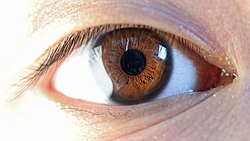
A limbal ring is a dark ring around the iris of the eye, where the sclera meets the cornea. [1] It is a dark-colored manifestation of the corneal limbus resulting from optical properties of the region. [2] The appearance and visibility of the limbal ring can be negatively affected by a variety of medical conditions concerning the peripheral cornea. [3] It has been suggested that limbal ring thickness may correlate with health or youthfulness and may contribute to facial attractiveness. [3] [4] The thickness of the limbal ring varies by pupil dilation - when the pupil is larger, the limbal ring narrows. [5] Some contact lenses are colored to simulate limbal rings. [1]

Contents

Youth, health, and attractiveness

Both health and age are positively correlated with a prominent limbal ring. [6] For instance, a darker limbal ring tends to be found more attractive than the absence of a limbal ring, suggesting that both sexes "use the limbal ring as a probabilistic indicator of reproductive fitness". [6] Furthermore, limbal rings appear to be most noticeable "for individuals relatively free from chronic health issues". [7]

Blue eye without a limbal ring Eye Central Heterochromia crop and lighter.jpg
Blue eye without a limbal ring

The limbal ring is thought to contain corneal epithelium stem cells. [8] Diseases involving a limbal stem cell deficiency are associated with blindness, as those with such deficiencies are "unable to maintain a stable corneal surface." [9] Transplantation of limbal stem cells is a promising therapy for limbal stem cell deficiency. [10]

See also

Related Research Articles

<span class="mw-page-title-main">Contact lens</span> Lenses placed on the eyes surface

Contact lenses, or simply contacts, are thin lenses placed directly on the surface of the eyes. Contact lenses are ocular prosthetic devices used by over 150 million people worldwide, and they can be worn to correct vision or for cosmetic or therapeutic reasons. In 2010, the worldwide market for contact lenses was estimated at $6.1 billion, while the US soft lens market was estimated at $2.1 billion. Multiple analysts estimated that the global market for contact lenses would reach $11.7 billion by 2015. As of 2010, the average age of contact lens wearers globally was 31 years old, and two-thirds of wearers were female.

<span class="mw-page-title-main">Keratoconus</span> Medical condition involving the eye

Keratoconus (KC) is a disorder of the eye that results in progressive thinning of the cornea. This may result in blurry vision, double vision, nearsightedness, irregular astigmatism, and light sensitivity leading to poor quality-of-life. Usually both eyes are affected. In more severe cases a scarring or a circle may be seen within the cornea.

<span class="mw-page-title-main">Cornea</span> Transparent front layer of the eye

The cornea is the transparent front part of the eye that covers the iris, pupil, and anterior chamber. Along with the anterior chamber and lens, the cornea refracts light, accounting for approximately two-thirds of the eye's total optical power. In humans, the refractive power of the cornea is approximately 43 dioptres. The cornea can be reshaped by surgical procedures such as LASIK.

<span class="mw-page-title-main">Phakic intraocular lens</span> Lens implanted in eye in addition to the natural lens

A phakic intraocular lens (PIOL) is a special kind of intraocular lens that is implanted surgically into the eye to correct myopia (nearsightedness). It is called "phakic" because the eye's natural lens is left untouched. Intraocular lenses that are implanted into eyes after the eye's natural lens has been removed during cataract surgery are known as pseudophakic.

<span class="mw-page-title-main">Corneal endothelium</span> Single layer of endothelial cells on the surface of the cornea

The corneal endothelium is a single layer of endothelial cells on the inner surface of the cornea. It faces the chamber formed between the cornea and the iris.

<span class="mw-page-title-main">Human eye</span> Sensory organ of vision

The human eye is a sensory organ, part of the sensory nervous system, that reacts to visible light and allows humans to use visual information for various purposes including seeing things, keeping balance, and maintaining circadian rhythm.

<span class="mw-page-title-main">Fuchs' dystrophy</span> Medical condition

Fuchs dystrophy, also referred to as Fuchs endothelial corneal dystrophy (FECD) and Fuchs endothelial dystrophy (FED), is a slowly progressing corneal dystrophy that usually affects both eyes and is slightly more common in women than in men. Although early signs of Fuchs dystrophy are sometimes seen in people in their 30s and 40s, the disease rarely affects vision until people reach their 50s and 60s.

<span class="mw-page-title-main">Recurrent corneal erosion</span> Medical condition

Recurrent corneal erosion is a disorder of the eyes characterized by the failure of the cornea's outermost layer of epithelial cells to attach to the underlying basement membrane. The condition is excruciatingly painful because the loss of these cells results in the exposure of sensitive corneal nerves. This condition can often leave patients with temporary blindness due to extreme light sensitivity (photophobia).

<span class="mw-page-title-main">Corneal cross-linking</span> Surgical procedure

Corneal cross-linking (CXL) with riboflavin (vitamin B2) and UV-A light is a surgical treatment for corneal ectasia such as keratoconus, PMD, and post-LASIK ectasia.

<span class="mw-page-title-main">Corneal dystrophy</span> Medical condition

Corneal dystrophy is a group of rare hereditary disorders characterised by bilateral abnormal deposition of substances in the transparent front part of the eye called the cornea.

<span class="mw-page-title-main">Stroma of cornea</span> Lamellated connective tissue of cornea

The stroma of the cornea is a fibrous, tough, unyielding, perfectly transparent and the thickest layer of the cornea of the eye. It is between Bowman's layer anteriorly, and Descemet's membrane posteriorly.

<span class="mw-page-title-main">Corneal neovascularization</span> Medical condition

Corneal neovascularization (CNV) is the in-growth of new blood vessels from the pericorneal plexus into avascular corneal tissue as a result of oxygen deprivation. Maintaining avascularity of the corneal stroma is an important aspect of corneal pathophysiology as it is required for corneal transparency and optimal vision. A decrease in corneal transparency causes visual acuity deterioration. Corneal tissue is avascular in nature and the presence of vascularization, which can be deep or superficial, is always pathologically related.

<span class="mw-page-title-main">Corneal ulcer</span> Medical condition of the eye

Corneal ulcer, also called keratitis, is an inflammatory or, more seriously, infective condition of the cornea involving disruption of its epithelial layer with involvement of the corneal stroma. It is a common condition in humans particularly in the tropics and in farming. In developing countries, children afflicted by vitamin A deficiency are at high risk for corneal ulcer and may become blind in both eyes persisting throughout life. In ophthalmology, a corneal ulcer usually refers to having an infection, while the term corneal abrasion refers more to a scratch injury.

<span class="mw-page-title-main">Corneal limbus</span> Border between the cornea and the sclera of the eye

The corneal limbus is the border between the cornea and the sclera. It contains limbal stem cells in its palisades of Vogt. It may be affected by cancer or aniridia, among other issues. The limbal ring is a visible dark ring around the iris of the eye composed of darkened areas of the corneal limbus.

<span class="mw-page-title-main">Corneal keratocyte</span>

Corneal keratocytes are specialized fibroblasts residing in the stroma. This corneal layer, representing about 85-90% of corneal thickness, is built up from highly regular collagenous lamellae and extracellular matrix components. Keratocytes play the major role in keeping it transparent, healing its wounds, and synthesizing its components. In the unperturbed cornea keratocytes stay dormant, coming into action after any kind of injury or inflammation. Some keratocytes underlying the site of injury, even a light one, undergo apoptosis immediately after the injury. Any glitch in the precisely orchestrated process of healing may cloud the cornea, while excessive keratocyte apoptosis may be a part of the pathological process in the degenerative corneal disorders such as keratoconus, and these considerations prompt the ongoing research into the function of these cells.

Peripheral Light Focusing (PLF) can be described as the focusing of Solar Ultraviolet Radiation (SUVR) at the nasal limbus of the cornea. SUVR incident at the temporal limbus passes across the corneal dome and focuses at the nasal limbus. The limbal region is a stem cell rich tissue. Ocular conditions such as pinguecula and pterygium may occur due to the result of this increased exposure to SUVR in limal area, but it is not the only cause of pterygium formation.

Post-LASIK ectasia is a condition similar to keratoconus where the cornea starts to bulge forwards at a variable time after LASIK, PRK, or SMILE corneal laser eye surgery. However, the physiological processes of post-LASIK ectasia seem to be different from keratoconus. The visible changes in the basal epithelial cell and anterior and posterior keratocytes linked with keratoconus were not observed in post-LASIK ectasia.

Corneal ectatic disorders or corneal ectasia are a group of uncommon, noninflammatory, eye disorders characterised by bilateral thinning of the central, paracentral, or peripheral cornea.

<span class="mw-page-title-main">Limbal stem cell</span>

Limbal stem cells, also known as corneal epithelial stem cells, are unipotent stem cells located in the basal epithelial layer of the corneal limbus. They form the border between the cornea and the sclera. Characteristics of limbal stem cells include a slow turnover rate, high proliferative potential, clonogenicity, expression of stem cell markers, as well as the ability to regenerate the entire corneal epithelium. Limbal stem cell proliferation has the role of maintaining the cornea; for example, by replacing cells that are lost via tears. Additionally, these cells also prevent the conjunctival epithelial cells from migrating onto the surface of the cornea.

Exposure keratopathy is medical condition affecting the cornea of eyes. It can lead to corneal ulceration and permanent loss of vision due to corneal opacity.

References

  1. 1 2 Johnson and Johnson Vision Care Inc. Tinted contact lenses with combined limbal ring and iris patterns. US7246903B2. United States Patent and Trademark Office, July 24, 2007. http://patft.uspto.gov/netacgi/nph-Parser?Sect1=PTO1&Sect2=HITOFF&p=1&u=/netahtml/PTO/srchnum.html&r=1&f=G&l=50&d=PALL&s1=7246903.PN.
  2. Shyu, Betsy P.; Wyatt, Harry J. (2009). "Appearance of the Human Eye: Optical Contributions to the 'Limbal Ring'". Optometry and Vision Science. 86 (9): E1069–77. doi:10.1097/OPX.0b013e3181b4f010. PMID   19648842. S2CID   39174174.
  3. 1 2 Peshek D, Semmaknejad N, Hoffman D, Foley P (2011). "Preliminary evidence that the limbal ring influences facial attractiveness" (PDF). Evolutionary Psychology. 9 (2): 137–146. doi: 10.1177/147470491100900201 . PMID   22947961.
  4. Brown M, Sacco DF (2018). "Put a (Limbal) Ring on It: Women Perceive Men's Limbal Rings as a Health Cue in Short-Term Mating Domains". Pers Soc Psychol Bull. 44 (1): 80–91. doi:10.1177/0146167217733072. PMID   28978250.
  5. Shyu, Betsy P.; Wyatt, Harry J. (September 2009). "Appearance of the Human Eye: Optical Contributions to the "Limbal Ring"". Optometry and Vision Science. 86 (9): E1069–E1077. doi:10.1097/OPX.0b013e3181b4f010. ISSN   1040-5488.
  6. 1 2 Peshek D, Semmaknejad N, Hoffman D, Foley P (2011). "Preliminary evidence that the limbal ring influences facial attractiveness" (PDF). Evolutionary Psychology. 9 (2): 137–146. doi: 10.1177/147470491100900201 . PMID   22947961.
  7. Brown M, Sacco DF (2018). "Put a (Limbal) Ring on It: Women Perceive Men's Limbal Rings as a Health Cue in Short-Term Mating Domains". Pers Soc Psychol Bull. 44 (1): 80–91. doi:10.1177/0146167217733072. PMID   28978250.
  8. Li, De-Quan; Chen, Zhuo; Song, Xiu Jun; de Paiva, Cintia S.; Kim, Hyun-Seung; Pflugfelder, Stephen C. (April 2005). "Partial enrichment of a population of human limbal epithelial cells with putative stem cell properties based on collagen type IV adhesiveness". Experimental Eye Research. 80 (4): 581–590. doi:10.1016/j.exer.2004.11.011. ISSN   0014-4835. PMC   2906384 . PMID   15781286.
  9. James, S. Elizabeth; Rowe, Andrea; Ilari, Luca; Daya, Sheraz; Martin, Robin (July 2001). "The Potential for Eye Bank Limbal Rings to Generate Cultured Corneal Epithelial Allografts". Cornea. 20 (5): 488–494. doi:10.1097/00003226-200107000-00010. ISSN   0277-3740. PMID   11413404. S2CID   21296292.
  10. Utheim, O. A.; Pasovic, L.; Raeder, S.; Eidet, J. R.; Fostad, I. G.; Sehic, A.; Roald, B.; Paz, M. F. de la; Lyberg, T.; Dartt, D. A.; Utheim, T. P. (2019-03-12). "Effects of explant size on epithelial outgrowth, thickness, stratification, ultrastructure and phenotype of cultured limbal epithelial cells". PLOS ONE. 14 (3): e0212524. Bibcode:2019PLoSO..1412524U. doi: 10.1371/journal.pone.0212524 . ISSN   1932-6203. PMC   6413940 . PMID   30861002.