Related Research Articles

Fibronectin is a high-molecular weight glycoprotein of the extracellular matrix that binds to membrane-spanning receptor proteins called integrins. It is approved for marketing as a topical solution in India by Central Drugs Standard Control organization in 2020 under the brand name FIBREGA for chronic wounds. Fibronectin also binds to other extracellular matrix proteins such as collagen, fibrin, and heparan sulfate proteoglycans.

The fluid mosaic model explains various characteristics regarding the structure of functional cell membranes. According to this biological model, there is a lipid bilayer in which protein molecules are embedded. The phospholipid bilayer gives fluidity and elasticity to the membrane. Small amounts of carbohydrates are also found in the cell membrane. The biological model, which was devised by Seymour Jonathan Singer and Garth L. Nicolson in 1972, describes the cell membrane as a two-dimensional liquid that restricts the lateral diffusion of membrane components. Such domains are defined by the existence of regions within the membrane with special lipid and protein cocoon that promote the formation of lipid rafts or protein and glycoprotein complexes. Another way to define membrane domains is the association of the lipid membrane with the cytoskeleton filaments and the extracellular matrix through membrane proteins. The current model describes important features relevant to many cellular processes, including: cell-cell signaling, apoptosis, cell division, membrane budding, and cell fusion. The fluid mosaic model is the most acceptable model of the plasma membrane. In this definition of the cell membrane, its main function is to act as a barrier between the contents inside the cell and the extracellular environment.

Nacre, also known as mother of pearl, is an organic–inorganic composite material produced by some molluscs as an inner shell layer. It is also the material of which pearls are composed. It is strong, resilient, and iridescent.
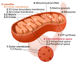
In the mitochondrion, the matrix is the space within the inner membrane. The word "matrix" stems from the fact that this space is viscous, compared to the relatively aqueous cytoplasm. The mitochondrial matrix contains the mitochondrial DNA, ribosomes, soluble enzymes, small organic molecules, nucleotide cofactors, and inorganic ions.[1] The enzymes in the matrix facilitate reactions responsible for the production of ATP, such as the citric acid cycle, oxidative phosphorylation, oxidation of pyruvate, and the beta oxidation of fatty acids.

In mammalian cells, vinculin is a membrane-cytoskeletal protein in focal adhesion plaques that is involved in linkage of integrin adhesion molecules to the actin cytoskeleton. Vinculin is a cytoskeletal protein associated with cell-cell and cell-matrix junctions, where it is thought to function as one of several interacting proteins involved in anchoring F-actin to the membrane.

An eggshell is the outer covering of a hard-shelled egg and of some forms of eggs with soft outer coats.

Mesenchyme is a type of loosely organized animal embryonic connective tissue of undifferentiated cells that give rise to most tissues, such as skin, blood or bone. The interactions between mesenchyme and epithelium help to form nearly every organ in the developing embryo.

Dystroglycan is a protein that in humans is encoded by the DAG1 gene.

Glycoprotein 130 is a transmembrane protein which is the founding member of the class of tall cytokine receptors. It forms one subunit of the type I cytokine receptor within the IL-6 receptor family. It is often referred to as the common gp130 subunit, and is important for signal transduction following cytokine engagement. As with other type I cytokine receptors, gp130 possesses a WSXWS amino acid motif that ensures correct protein folding and ligand binding. It interacts with Janus kinases to elicit an intracellular signal following receptor interaction with its ligand. Structurally, gp130 is composed of five fibronectin type-III domains and one immunoglobulin-like C2-type (immunoglobulin-like) domain in its extracellular portion.

Mitochondrial membrane transport proteins, also known as mitochondrial carrier proteins, are proteins which exist in the membranes of mitochondria. They serve to transport molecules and other factors, such as ions, into or out of the organelles. Mitochondria contain both an inner and outer membrane, separated by the inter-membrane space, or inner boundary membrane. The outer membrane is porous, whereas the inner membrane restricts the movement of all molecules. The two membranes also vary in membrane potential and pH. These factors play a role in the function of mitochondrial membrane transport proteins. There are 53 discovered human mitochondrial membrane transporters, with many others that are known to still need discovered.
Fibroin is an insoluble protein present in silk produced by numerous insects, such as the larvae of Bombyx mori, and other moth genera such as Antheraea, Cricula, Samia and Gonometa. Silk in its raw state consists of two main proteins, sericin and fibroin, with a glue-like layer of sericin coating two singular filaments of fibroin called brins. Silk fibroin is considered a β-keratin related to proteins that form hair, skin, nails and connective tissues.

Nuclear bodies are membraneless structures found in the cell nuclei of eukaryotic cells. Nuclear bodies include Cajal bodies, the nucleolus, and promyelocytic leukemia protein (PML) nuclear bodies. Nuclear bodies also include ND10s. ND stands for nuclear domain, and 10 refers to the number of dots seen.

FBLN1 is the gene encoding fibulin-1, an extracellular matrix and plasma protein.

Syndecan-4 is a protein that in humans is encoded by the SDC4 gene. Syndecan-4 is one of the four vertebrate syndecans and has a molecular weight of ~20 kDa. Syndecans are the best-characterized plasma membrane proteoglycans. Their intracellular domain of membrane-spanning core protein interacts with actin cytoskeleton and signaling molecules in the cell cortex. Syndecans are normally found on the cell surface of fibroblasts and epithelial cells. Syndecans interact with fibronectin on the cell surface, cytoskeletal and signaling proteins inside the cell to modulate the function of integrin in cell-matrix adhesion. Also, syndecans bind to FGFs and bring them to the FGF receptor on the same cell. As a co-receptor or regulator, mutated certain proteoglycans could cause severe developmental defects, like disordered distribution or inactivation of signaling molecules.

Collagen alpha-3(IV) chain is a protein that in humans is encoded by the COL4A3 gene.

Cytoglobin is the protein product of CYGB, a human and mammalian gene.

The molluscshell is typically a calcareous exoskeleton which encloses, supports and protects the soft parts of an animal in the phylum Mollusca, which includes snails, clams, tusk shells, and several other classes. Not all shelled molluscs live in the sea; many live on the land and in freshwater.

Mollusca is the second-largest phylum of invertebrate animals, after Arthropoda; members are known as molluscs or mollusks. Around 76,000 extant species of molluscs are recognized. The number of fossil species is estimated between 60,000 and 100,000 additional species. The proportion of undescribed species is very high. Many taxa remain poorly studied.

Mineralized tissues are biological tissues that incorporate minerals into soft matrices. Typically these tissues form a protective shield or structural support. Bone, mollusc shells, deep sea sponge Euplectella species, radiolarians, diatoms, antler bone, tendon, cartilage, tooth enamel and dentin are some examples of mineralized tissues.
Aspein is an unusually acidic bivalve shell matrix protein, which may have important roles in calcium carbonate biomineralization. The Aspein gene encodes a sequence of 413 amino acids, including a high proportion of Asp (60.4%), Gly (16.0%), and Ser (13.2%), and the predicted isoelectric point is 1.78. This is the most acidic of all the molluscan shell matrix proteins sequenced so far, or probably even of all known proteins on earth. The main body of aspein is occupied by (Asp)2–10 sequences punctuated with Ser–Gly dipeptides. RT-PCR demonstrated that the transcript of aspein is expressed at the outer edge of the mantle, corresponding to the calcitic prismatic layer, but not at the inner part of the mantle, corresponding to the aragonitic nacreous layer. Aspein is likely responsible for directed formation of calcite in the shell of the pearl oyster Pinctada fucata.
References
- 1 2 Shen, X.; Belcher, A. M.; Hansma, P. K.; Stucky, G. D.; Morse, D. E. (1997). "Molecular Cloning and Characterization of Lustrin A, a Matrix Protein from Shell and Pearl Nacre of Haliotis rufescens". Journal of Biological Chemistry. 272 (51): 32472–81. doi: 10.1074/jbc.272.51.32472 . PMID 9405458.
- ↑ Jackson, D.; McDougall, C.; Green, K.; Simpson, F.; Wörheide, G.; Degnan, B. (2006). "A rapidly evolving secretome builds and patterns a sea shell". BMC Biology. 4: 40. doi: 10.1186/1741-7007-4-40 . PMC 1676022 . PMID 17121673.
- 1 2 Marin, F.; Luquet, G. (2004). "Molluscan shell proteins". Comptes Rendus Palevol. 3 (6–7): 469–492. doi:10.1016/j.crpv.2004.07.009.