Related Research Articles

Neurofibromatosis (NF) is a group of three conditions in which tumors grow in the nervous system. The three types are neurofibromatosis type I (NF1), neurofibromatosis type II (NF2), and schwannomatosis. In NF1 symptoms include light brown spots on the skin, freckles in the armpit and groin, small bumps within nerves, and scoliosis. In NF2, there may be hearing loss, cataracts at a young age, balance problems, flesh colored skin flaps, and muscle wasting. In schwannomatosis there may be pain either in one location or in wide areas of the body. The tumors in NF are generally non-cancerous.

Proteus syndrome is a rare disorder with a genetic background that can cause tissue overgrowth involving all three embryonic lineages. Patients with Proteus syndrome tend to have an increased risk of embryonic tumor development. The clinical and radiographic symptoms of Proteus syndrome are highly variable, as are its orthopedic manifestations.

Osteosclerosis is a disorder that is characterized by abnormal hardening of bone and an elevation in bone density. It may predominantly affect the medullary portion and/or cortex of bone. Plain radiographs are a valuable tool for detecting and classifying osteosclerotic disorders. It can manifest in localized or generalized osteosclerosis. Localized osteosclerosis can be caused by Legg–Calvé–Perthes disease, sickle-cell disease and osteoarthritis among others. Osteosclerosis can be classified in accordance with the causative factor into acquired and hereditary.

Genu valgum, commonly called "knock-knee", is a condition in which the knees angle in and touch each other when the legs are straightened. Individuals with severe valgus deformities are typically unable to touch their feet together while simultaneously straightening the legs. The term originates from the Latin genu, 'knee', and valgus which means "bent outwards", but is also used to describe the distal portion of the knee joint which bends outwards and thus the proximal portion seems to be bent inwards.

Genu varum is a varus deformity marked by (outward) bowing at the knee, which means that the lower leg is angled inward (medially) in relation to the thigh's axis, giving the limb overall the appearance of an archer's bow. Usually medial angulation of both lower limb bones is involved.

Local gigantism or localised gigantism is a condition in which a certain part of the body acquires larger than normal size due to excessive growth of the anatomical structures or abnormal accumulation of substances. It is more common in fingers and toes, where it is termed macrodactyly. However, sometimes an entire limb may be enlarged.
An osteochondrodysplasia, or skeletal dysplasia, is a disorder of the development of bone and cartilage. Osteochondrodysplasias are rare diseases. About 1 in 5,000 babies are born with some type of skeletal dysplasia. Nonetheless, if taken collectively, genetic skeletal dysplasias or osteochondrodysplasias comprise a recognizable group of genetically determined disorders with generalized skeletal affection. These disorders lead to disproportionate short stature and bone abnormalities, particularly in the arms, legs, and spine. Skeletal dysplasia can result in marked functional limitation and even mortality.

The epiphyseal plate is a hyaline cartilage plate in the metaphysis at each end of a long bone. It is the part of a long bone where new bone growth takes place; that is, the whole bone is alive, with maintenance remodeling throughout its existing bone tissue, but the growth plate is the place where the long bone grows longer.

Pseudoachondroplasia is an inherited disorder of bone growth. It is a genetic autosomal dominant disorder. It is generally not discovered until 2–3 years of age, since growth is normal at first. Pseudoachondroplasia is usually first detected by a drop of linear growth in contrast to peers, a waddling gait or arising lower limb deformities.

Blount's disease is a growth disorder of the tibia which causes the lower leg to angle inward, resembling a bowleg. It is also known as "tibia vara".
Overgrowth syndromes in children constitute a group of rare disorders that are characterised by tissue hypertrophy. Individual overgrowth syndromes have been shown to overlap with regard to clinical and radiologic features. The details of the genetic bases of these syndromes are unfolding. Any of the three embryonic tissue layers may be involved. The syndromes may manifest in localized or generalized tissue overgrowth. Latitudinal and longitudinal growth may be affected. Nevertheless, the musculoskeletal features are central to the diagnosis of some syndromes such as Proteus syndrome.

Winchester syndrome is a rare hereditary connective tissue disease described in 1969, of which the main characteristics are short stature, marked contractures of joints, opacities in the cornea, coarse facial features, dissolution of the carpal and tarsal bones, and osteoporosis. Winchester syndrome was once considered to be related to a similar condition, multicentric osteolysis, nodulosis, and arthropathy (MONA). However, it was discovered that the two are caused by mutations found in different genes; however they mostly produce the same phenotype or clinical picture. Appearances resemble rheumatoid arthritis. Increased uronic acid is demonstrated in cultured fibroblasts from the skin and to a lesser degree in both parents. Despite initial tests not showing increased mucopolysaccharide excretion, the disease was regarded as a mucopolysaccharidosis. Winchester syndrome is thought to be inherited as an autosomal recessive trait.

Toddler's fractures are bone fractures of the distal (lower) part of the shin bone (tibia) in toddlers and other young children. The fracture is found in the distal two thirds of the tibia in 95% of cases, is undisplaced and has a spiral pattern. It occurs after low-energy trauma, sometimes with a rotational component.

Klippel–Trénaunay syndrome, formerly Klippel–Trénaunay–Weber syndrome and sometimes angioosteohypertrophy syndrome and hemangiectatic hypertrophy, is a rare congenital medical condition in which blood vessels and/or lymph vessels fail to form properly. The three main features are nevus flammeus, venous and lymphatic malformations, and soft-tissue hypertrophy of the affected limb. It is similar to, though distinctly separate from, the less common Parkes Weber syndrome.

Parkes Weber syndrome (PWS) is a congenital disorder of the vascular system. It is an extremely rare condition, and its exact prevalence is unknown. It is named after British dermatologist Frederick Parkes Weber, who first described the syndrome in 1907.

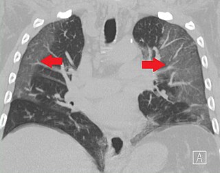
Ground-glass opacity (GGO) is a finding seen on chest x-ray (radiograph) or computed tomography (CT) imaging of the lungs. It is typically defined as an area of hazy opacification (x-ray) or increased attenuation (CT) due to air displacement by fluid, airway collapse, fibrosis, or a neoplastic process. When a substance other than air fills an area of the lung it increases that area's density. On both x-ray and CT, this appears more grey or hazy as opposed to the normally dark-appearing lungs. Although it can sometimes be seen in normal lungs, common pathologic causes include infections, interstitial lung disease, and pulmonary edema.

A cavernous liver hemangioma or hepatic hemangioma is a benign tumor of the liver composed of hepatic endothelial cells. It is the most common benign liver tumour, and is usually asymptomatic and diagnosed incidentally on radiological imaging. Liver hemangiomas are thought to be congenital in origin. Several subtypes exist, including the giant hepatic haemangioma, which can cause significant complications.

Adenomyomatosis is a benign condition characterized by hyperplastic changes of unknown cause involving the wall of the gallbladder. Adenomyomatosis is caused by an overgrowth of the mucosa, thickening of the muscular wall, and formation of intramural diverticula or sinus tracts termed Rokitansky–Aschoff sinuses, also called entrapped epithelial crypts.

Malignant infantile osteopetrosis is a rare osteosclerosing type of skeletal dysplasia that typically presents in infancy and is characterized by a unique radiographic appearance of generalized hyperostosis.
PIK3CA-related overgrowth spectrum (PROS) is an umbrella term for rare syndromes characterized by malformations and tissue overgrowth caused by somatic mutations in PIK3CA gene. In PROS diseases individuals malformations are seen in several different tissues such as skin, vasculature, bones, fat and brain tissue depending on the specific disease.
References
- ↑ Prasetyono TO, Hanafi E, Astriana W (2015). "A Review of Macrodystrophia Lipomatosa: Revisitation". Archives of Plastic Surgery. 42 (4): 391–406. doi: 10.5999/aps.2015.42.4.391 . PMC 4513046 . PMID 26217558.
{{cite journal}}: CS1 maint: multiple names: authors list (link) - 1 2 3 4 Abdulhady, H; El-Sobky, TA; Elsayed, NS; Sakr, HM (11 June 2018). "Clinical and imaging features of pedal macrodystrophia lipomatosa in two children with differential diagnosis review". Journal of Musculoskeletal Surgery and Research. 2 (3): 130. doi: 10.4103/jmsr.jmsr_8_18 . S2CID 80970016.
- ↑ EL-Sobky TA, Elsayed SM, EL Mikkawy DME (2015). "Orthopaedic manifestations of Proteus syndrome in a child with literature update". Bone Rep. 3: 104–108. doi:10.1016/j.bonr.2015.09.004. PMC 5365241 . PMID 28377973.
{{cite journal}}: CS1 maint: multiple names: authors list (link) - ↑ Friedman, JM (11 January 2018). "Neurofibromatosis 1". GeneReviews. University of Washington, Seattle. PMID 20301288 . Retrieved 30 April 2018.
- ↑ Sung, HM; Chung, HY; Lee, SJ; et, al (2015). "Clinical experience of the Klippel-Trenaunay syndrome". Arch Plast Surg. 42 (5): 552–8. doi:10.5999/aps.2015.42.5.552. PMC 4579165 . PMID 26430625.
- ↑ Nguyen, TA; Krakowski, AC; Naheedy, JH; Kruk, PG; Friedlander, SF (2015). "Imaging Pediatric Vascular Lesions". Clin Aesthet Dermatol. 8 (12): 27–41. PMC 4689509 . PMID 26705446.
- Bailey EJ, Thompson FM, Bohne W, Dyal C (Feb 1997). "Macrodystrophia lipomatosa of the foot: a report of three cases and literature review". Foot Ankle Int. 18 (2): 89–93. doi:10.1177/107110079701800209. PMID 9043881. S2CID 31353649.
{{cite journal}}: CS1 maint: multiple names: authors list (link) - Goldman AB, Kaye JJ (1977). "Macrodystrophia Lipomatosa: Radiographic Diagnosis". Am. J. Roentgenol. 128 (1): 101–105. doi:10.2214/ajr.128.1.101. PMID 401563.
- Blacksin M., Barnes FJ, Lyons MM (1992). "MR Diagnosis of Macrodystrophia Lipomatosa". Am. J. Roentgenol. 158 (6): 1295–1297. doi: 10.2214/ajr.158.6.1590127 . PMID 1590127.
{{cite journal}}: CS1 maint: multiple names: authors list (link)