
Geophysics is a subject of natural science concerned with the physical processes and physical properties of the Earth and its surrounding space environment, and the use of quantitative methods for their analysis. Geophysicists, who usually study geophysics, physics, or one of the Earth sciences at the graduate level, complete investigations across a wide range of scientific disciplines. The term geophysics classically refers to solid earth applications: Earth's shape; its gravitational, magnetic fields, and electromagnetic fields ; its internal structure and composition; its dynamics and their surface expression in plate tectonics, the generation of magmas, volcanism and rock formation. However, modern geophysics organizations and pure scientists use a broader definition that includes the water cycle including snow and ice; fluid dynamics of the oceans and the atmosphere; electricity and magnetism in the ionosphere and magnetosphere and solar-terrestrial physics; and analogous problems associated with the Moon and other planets.
Medical physics deals with the application of the concepts and methods of physics to the prevention, diagnosis and treatment of human diseases with a specific goal of improving human health and well-being. Since 2008, medical physics has been included as a health profession according to International Standard Classification of Occupation of the International Labour Organization.

Tomography is imaging by sections or sectioning that uses any kind of penetrating wave. The method is used in radiology, archaeology, biology, atmospheric science, geophysics, oceanography, plasma physics, materials science, cosmochemistry, astrophysics, quantum information, and other areas of science. The word tomography is derived from Ancient Greek τόμος tomos, "slice, section" and γράφω graphō, "to write" or, in this context as well, "to describe." A device used in tomography is called a tomograph, while the image produced is a tomogram.

Electrical impedance tomography (EIT) is a noninvasive type of medical imaging in which the electrical conductivity, permittivity, and impedance of a part of the body is inferred from surface electrode measurements and used to form a tomographic image of that part. Electrical conductivity varies considerably among various biological tissues or the movement of fluids and gases within tissues. The majority of EIT systems apply small alternating currents at a single frequency, however, some EIT systems use multiple frequencies to better differentiate between normal and suspected abnormal tissue within the same organ.
Microwave spectroscopy is the spectroscopy method that employs microwaves, i.e. electromagnetic radiation at GHz frequencies, for the study of matter.

Magnetotellurics (MT) is an electromagnetic geophysical method for inferring the earth's subsurface electrical conductivity from measurements of natural geomagnetic and geoelectric field variation at the Earth's surface.
The Robinson oscillator is an electronic oscillator circuit originally devised for use in the field of continuous wave (CW) nuclear magnetic resonance (NMR). It was a development of the marginal oscillator. Strictly one should distinguish between the marginal oscillator and the Robinson oscillator, although sometimes they are conflated and referred to as a Robinson marginal oscillator. Modern magnetic resonance imaging (MRI) systems are based on pulsed NMR; they do not rely on the use of such oscillators.

Electrical capacitance tomography (ECT) is a method for determination of the dielectric permittivity distribution in the interior of an object from external capacitance measurements. It is a close relative of electrical impedance tomography and is proposed as a method for industrial process monitoring.

A biosignal is any signal in living beings that can be continually measured and monitored. The term biosignal is often used to refer to bioelectrical signals, but it may refer to both electrical and non-electrical signals. The usual understanding is to refer only to time-varying signals, although spatial parameter variations are sometimes subsumed as well.

Feature-oriented scanning (FOS) is a method of precision measurement of surface topography with a scanning probe microscope in which surface features (objects) are used as reference points for microscope probe attachment. With FOS method, by passing from one surface feature to another located nearby, the relative distance between the features and the feature neighborhood topographies are measured. This approach allows to scan an intended area of a surface by parts and then reconstruct the whole image from the obtained fragments. Beside the mentioned, it is acceptable to use another name for the method – object-oriented scanning (OOS).

Process tomography consists of tomographic imaging of systems, such as process pipes in industry. In tomography the 3D distribution of some physical quantity in the object is determined. There is a widespread need to get tomographic information about process. This information can be used, for example, in the design and control of processes.
Transient electromagnetics,, is a geophysical exploration technique in which electric and magnetic fields are induced by transient pulses of electric current and the subsequent decay response measured. TEM / TDEM methods are generally able to determine subsurface electrical properties, but are also sensitive to subsurface magnetic properties in applications like UXO detection and characterization. TEM/TDEM surveys are a very common surface EM technique for mineral exploration, groundwater exploration, and for environmental mapping, used throughout the world in both onshore and offshore applications.
Frequency scanning interferometry (FSI) is an absolute distance measurement technique, for measuring the distance between a pair of points, along a line-of-sight. The power of the FSI technique lies in its ability to make many such distance measurements, simultaneously.

Scanning thermal microscopy (SThM) is a type of scanning probe microscopy that maps the local temperature and thermal conductivity of an interface. The probe in a scanning thermal microscope is sensitive to local temperatures – providing a nano-scale thermometer. Thermal measurements at the nanometer scale are of both scientific and industrial interest. The technique was invented by Clayton C. Williams and H. Kumar Wickramasinghe in 1986.
Electrical impedance myography, or EIM, is a non-invasive technique for the assessment of muscle health that is based on the measurement of the electrical impedance characteristics of individual muscles or groups of muscles. The technique has been used for the purpose of evaluating neuromuscular diseases both for their diagnosis and for their ongoing assessment of progression or with therapeutic intervention. Muscle composition and microscopic structure change with disease, and EIM measures alterations in impedance that occur as a result of disease pathology. EIM has been specifically recognized for its potential as an ALS biomarker by Prize4Life, a 501(c)(3) nonprofit organization dedicated to accelerating the discovery of treatments and cures for ALS. The $1M ALS Biomarker Challenge focused on identifying a biomarker precise and reliable enough to cut Phase II drug trials in half. The prize was awarded to Dr. Seward Rutkove, chief, Division of Neuromuscular Disease, in the Department of Neurology at Beth Israel Deaconess Medical Center and Professor of Neurology at Harvard Medical School, for his work in developing the technique of EIM and its specific application to ALS. It is hoped that EIM as a biomarker will result in the more rapid and efficient identification of new treatments for ALS. EIM has shown sensitivity to disease status in a variety of neuromuscular conditions, including radiculopathy, inflammatory myopathy, Duchenne muscular dystrophy, and spinal muscular atrophy.

A fluid dynamic gauge (FDG) is a measurement technique used to study the behaviour of soft deposit layers in a liquid environment. It employs fluid mechanics to determine the thickness of the layer, and can also be used to obtain a measure of its strength. It was inspired by the technique of pneumatic gauging, which relies on a flow of air rather than the process liquid. Fluid dynamic gauging can be conducted as an in-line measuring technique, but is more commonly used as a research tool.

Near-surface geophysics is the use of geophysical methods to investigate small-scale features in the shallow subsurface. It is closely related to applied geophysics or exploration geophysics. Methods used include seismic refraction and reflection, gravity, magnetic, electric, and electromagnetic methods. Many of these methods were developed for oil and mineral exploration but are now used for a great variety of applications, including archaeology, environmental science, forensic science, military intelligence, geotechnical investigation, treasure hunting, and hydrogeology. In addition to the practical applications, near-surface geophysics includes the study of biogeochemical cycles.
Lorentz force velocimetry (LFV) is a noncontact electromagnetic flow measurement technique. LFV is particularly suited for the measurement of velocities in liquid metals like steel or aluminium and is currently under development for metallurgical applications. The measurement of flow velocities in hot and aggressive liquids such as liquid aluminium and molten glass constitutes one of the grand challenges of industrial fluid mechanics. Apart from liquids, LFV can also be used to measure the velocity of solid materials as well as for detection of micro-defects in their structures.
EIDORS is an open-source software tool box written mainly in MATLAB/GNU Octave designed primarily for image reconstruction from electrical impedance tomography (EIT) data, in a biomedical, industrial or geophysical setting. The name was originally an acronym for Electrical Impedance Tomography and Diffuse Optical Reconstruction Software. While the name reflects the original intention to cover image reconstruction of data from the mathematically similar near infra red diffuse optical imaging, to date there has been little development in that area.
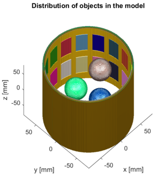
Three-dimensional electrical capacitance tomography also known as electrical capacitance volume tomography (ECVT) is a non-invasive 3D imaging technology applied primarily to multiphase flows. It was introduced in the early 2000s as an extension of the conventional two-dimensional ECT. In conventional electrical capacitance tomography, sensor plates are distributed around a surface of interest. Measured capacitance between plate combinations is used to reconstruct 2D images (tomograms) of material distribution. Because the ECT sensor plates are required to have lengths on the order of the domain cross-section, 2D ECT does not provide the required resolution in the axial dimension. In ECT, the fringing field from the edges of the plates is viewed as a source of distortion to the final reconstructed image and is thus mitigated by guard electrodes. 3D ECT exploits this fringing field and expands it through 3D sensor designs that deliberately establish an electric field variation in all three dimensions. In 3D tomography, the data are acquired in 3D geometry, and the reconstruction algorithm produces the three-dimensional image directly, in contrast to 2D tomography, where 3D information might be obtained by stacking 2D slices reconstructed individually.